Right Frontoparietal Craniotomy: A Case Study of Successful Brain Tumor Removal in a 48-Y-O Female
PACE Hospitals
PACE Hospitals' Expert Neurosurgery Team successfully performed a right frontoparietal craniotomy followed by tumor excision on a 48-year-old female patient who presented with seizures and weakness in the left upper and lower limbs. The procedure aimed to achieve complete tumor removal, relieve neurological symptoms, and enhance the patient’s overall functional recovery and quality of life.
Chief Complaints
A 48-year-old female patient, with a
BMI of 23.4, presented to the Neurosurgery Department at
PACE Hospitals, Hitech City, Hyderabad, with complaints of
seizures and weakness in the left upper and lower limbs for the past month. Her symptoms had gradually worsened, significantly affecting her daily activities and prompting further neurological evaluation.
History of Present Illness
The patient experienced the onset of seizures approximately one month ago, followed by progressive weakness in the left upper and lower limbs. She underwent initial evaluation at an outside facility, where imaging revealed a right fronto-parietal parasagittal space-occupying lesion (SOL). Seeking specialised care, she presented to PACE Hospitals for further evaluation and definitive management.
Past Medical History
She had a known history of hypertension and hypothyroidism, for which she had been on regular medication. Both conditions were well-controlled at the time of presentation.
General Examination
Upon admission to PACE Hospitals, the patient was hemodynamically stable. General examination revealed that she was moderately built and nourished, with no signs of systemic illness. Cardiovascular and respiratory examinations were unremarkable, with normal heart sounds (S1, S2) and adequate bilateral air entry. Abdominal examination was normal, with a soft and non-tender abdomen. However, neurological evaluation indicated mild motor weakness in the left upper and lower limbs, with muscle strength graded at 4/5, while the right side showed normal strength (5/5). Bilateral flexor plantar reflexes and deep tendon reflexes graded at 2+ were noted, supporting the presence of a focal neurological deficit consistent with a right fronto-parietal lesion.
Diagnosis
As the patient had arrived with a preliminary diagnosis based on prior neuroimaging from another centre, the Neurosurgery team at PACE Hospitals carefully evaluated all existing reports, including MRI scans and neurological assessments.
Given her presenting symptoms of seizures and left-sided weakness, along with imaging performed at an outside facility suggesting a right Fronto-Parietal Parasagittal Space-Occupying Lesion (SOL), an intracranial tumor was strongly suspected.
To ensure diagnostic precision and guide appropriate surgical planning, the team initiated a comprehensive reassessment using a multidisciplinary approach, incorporating advanced neuroimaging and neurosurgical expertise.
To support the diagnosis and assess the extent of the condition, a series of relevant investigations were conducted:
MRI Brain: A contrast-enhanced brain MRI was performed as the first-line imaging modality. It revealed a well-defined, enhancing lesion in the right fronto-parietal parasagittal region, consistent with features typically seen in meningiomas, such as a dural tail sign and strong homogeneous enhancement. The MRI provided critical information regarding the tumor’s size, precise location, vascularity, and its relationship with adjacent brain structures.
CT Scan Brain: A CT scan of the brain was also conducted to assess any associated calcifications and to evaluate potential involvement of the overlying skull. The scan confirmed hyperdense areas within the lesion and helped rule out any acute haemorrhagic or bony complications, which is especially important for surgical planning.
Functional MRI (fMRI) and Diffusion Tensor Imaging (DTI): Given the tumor’s proximity to the motor cortex, advanced imaging with fMRI and DTI was performed. These modalities helped map the functional areas of the brain and the surrounding white matter tracts, ensuring that surgical planning prioritized the preservation of motor functions and minimized neurological risks.
Magnetic Resonance Spectroscopy (MRS): MRS was used to assess the metabolic profile of the lesion. The findings were consistent with a benign tumor, most likely a meningioma, further supporting the radiological diagnosis and ruling out high-grade malignancy.
Based on the confirmed diagnosis, the patient was advised to undergo Brain Tumor Treatment in Hyderabad, India, under the expert care of the Neurosurgery Department.
Based on the findings from the imaging and the outside reports, the Neurosurgery team meticulously reviewed all the data. A comprehensive reassessment was done, and a detailed surgical plan was prepared to ensure a precise and safe tumor removal, prioritizing the preservation of neurological function.
Medical Decision-Making (MDM)
Following her admission to PACE Hospitals, the patient was evaluated in detail by Dr. UL Sandeep Varma, Consultant Neurosurgeon. A comprehensive assessment of her clinical condition, along with a thorough review of imaging findings, was carried out. Based on the neurological deficits and the location of the lesion, the multidisciplinary medical team concluded that the most appropriate course of action would be to perform a right fronto-parietal craniotomy with tumor excision under general anaesthesia. This surgical approach was planned to relieve her neurological symptoms, restore motor function, and prevent further progression of the lesion.
Surgical Procedure
Following the decision, the patient was scheduled for a Right Frontoparietal Craniotomy and Tumor Excision Surgery in Hyderabad at PACE Hospitals, under the expert supervision of the Neurosurgery Department.
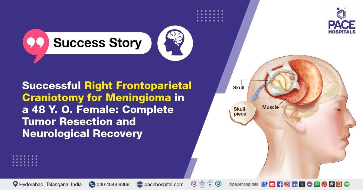
A right frontoparietal craniotomy is a neurosurgical procedure that involves temporarily removing a portion of the skull to reach the tumor, which is then carefully excised under microscopic guidance. This procedure is typically performed under general anaesthesia and guided by preoperative imaging to minimise damage to healthy brain tissue. It is commonly used for space-occupying lesions like meningiomas, gliomas, or metastases, particularly when they cause neurological symptoms like seizures or limb weakness.
After receiving anaesthesia clearance, the patient was taken to the operating room, where she underwent a right frontoparietal craniotomy under general anaesthesia. The procedure began with careful painting and draping of the surgical area, followed by an incision over the right frontoparietal region. Multiple burr holes were drilled, and the bone flap was elevated to gain access to the underlying tumor.
Once the tumor was located, it was meticulously excised in a piecemeal fashion, ensuring minimal disruption to the surrounding brain tissue while preserving critical neurovascular structures. A small rim of tissue was left along the draining cortical veins to prevent vascular injury.
Haemostasis was achieved successfully, and a lax duroplasty was performed using pericranium to reinforce the dural closure. The bone flap was carefully repositioned, and the surgical site was closed in layers, with a surgical drain placed to prevent any postoperative fluid accumulation.
Histopathological Examination: Following surgical excision of the tumor via craniotomy, tissue was sent for histopathological analysis. This definitive diagnostic step confirmed the tumor type, grade and ruled out any atypical or malignant transformation.
The procedure was completed without any immediate complications, and the patient was closely monitored in the postoperative period for recovery.
Postoperative Care
The postoperative care plan focused on ensuring a smooth recovery, closely monitoring for any complications, and planning further treatment based on the histopathological findings. Following the surgery, the patient was shifted to the ICU for close observation. On postoperative examination, the surgical wound was found to be healthy, with no signs of infection or complications. The patient’s vital signs remained stable throughout the recovery period. Neurological assessment showed improved motor strength, with power graded at 4+/5 in the left upper and lower limbs—indicating partial recovery—and 5/5 in the right upper and lower limbs, consistent with normal function. These findings reflected encouraging postoperative progress in line with the surgical objectives.
Her postoperative course was smooth and uneventful, with stable vital signs and no new neurological deficits. As her condition remained stable and the surgical site showed healthy healing, the neurosurgical team successfully removed the surgical drain on the second postoperative day. With steady neurological improvement and no signs of complications, she was prepared for discharge with appropriate post-discharge care instructions and follow-up plans
Discharge Notes
As the patient showed good postoperative progress, the medical team proceeded with discharge in a hemodynamically stable condition. She was given comprehensive instructions for continued care, including dietary modifications appropriate to her recovery status, proper wound care, and scheduled follow-up appointments to monitor her progress. The patient was advised to strictly adhere to the prescribed medications, maintain wound hygiene, engage in simple daily activities, and avoid any strenuous exercises to support a safe and smooth recovery.
Discharge Medications
Upon discharge, the patient was prescribed a tailored combination of medications to support her recovery and prevent complications. Antibiotics were given to prevent postoperative infections, while proton pump inhibitors (PPIs) were prescribed to protect the gastric lining from medication-induced irritation. Anticonvulsants were continued to manage and prevent seizures, and cerebroactive drugs were prescribed to enhance neurological recovery. Additionally, nutritional supplements, calcium channel blockers (for hypertension), analgesics for pain relief, laxatives to prevent constipation due to reduced mobility, and other medications were included in the regimen.
Emergency Care
The patient was informed to contact the Emergency ward at PACE Hospitals in case of any emergency or development of symptoms like seizures, fever, loss of consciousness (LOC), weakness of limbs, or vomiting.
Review and Follow-Up Notes
The patient was advised to schedule a follow-up appointment with the Neurosurgeon in Hyderabad at PACE Hospitals after 10 days. During this visit, the staples were planned to be removed, and her recovery was to be assessed for further management.
Conclusion
This case highlights the effectiveness of craniotomy as a key approach in Meningioma Treatment in Hyderabad, India, allowing for successful tumour removal while preserving the integrity of surrounding brain tissue. The procedure facilitated a faster recovery and ensured the protection of vital neurological structures, demonstrating the advanced neurosurgical expertise available in the region.
The Importance of Craniotomy in Treating Parasagittal Meningioma
Craniotomy plays a crucial role in the surgical management of brain tumors, particularly when the tumor is large, located near critical brain areas, or involves vascular structures. Open surgery allows neurosurgeons direct access to the lesion, enabling precise tumor excision while preserving surrounding healthy brain tissue and vital neurovascular structures. While minimally invasive procedures can be beneficial in select cases, they may not offer sufficient visibility or control for complex or deeply situated tumors like parasagittal meningiomas.
Large tumors often require piecemeal removal, which is more effectively managed through open craniotomy. This method also allows for intraoperative brain mapping and monitoring, reducing the risk of postoperative neurological deficits. In the case of this patient, the neurosurgeon/neurosurgery doctor carefully assessed her clinical condition, including seizures and left-sided weakness, along with imaging that showed a right fronto-parietal parasagittal meningioma close to critical motor areas. Given the size, location, and proximity to draining cortical veins, the neurosurgeon determined that craniotomy was the safest and most effective approach to excise the tumor while preserving essential brain structures. Minimally invasive techniques were deemed unsuitable due to the complex positioning of the lesion and the need for precise dissection of delicate neurovascular tissues. Therefore, craniotomy provided the best opportunity for complete tumor removal and optimal neurological recovery.
Share on
Request an appointment
Fill in the appointment form or call us instantly to book a confirmed appointment with our super specialist at 04048486868
Appointment request - health articles
Recent Articles


