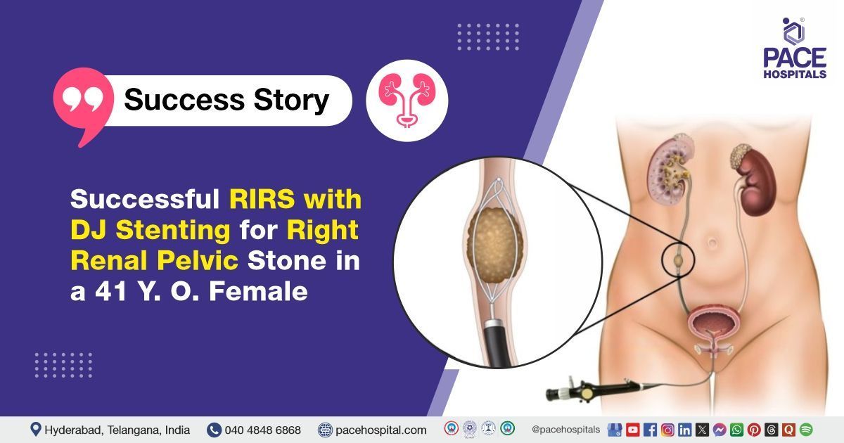
Successful RIRS with DJ Stenting for Right Renal Pelvic Stone in a 41-Year-Old Female
PACE Hospitals
The Urology team at PACE Hospitals successfully performed Right Retrograde Intrarenal Surgery (RIRS) with DJ stenting in a 41-year-old female who presented with complaints of right flank pain. Imaging revealed a 14 × 7 mm calculus in the right renal pelvis, causing discomfort and potential obstruction. The procedure aimed to remove the renal stone, thereby relieving the obstruction, alleviating symptoms, and promoting optimal kidney function and healing.
Chief Complaints
A 41-year-old female with a BMI of 23.4 presented to the Department of Urology at PACE Hospitals, Hitech City, Hyderabad, with complaints of persistent right flank pain that had been ongoing for several days. The pain was described as dull, aching and moderate in intensity.
She did not report any associated urinary symptoms such as dysuria, haematuria, nausea, or vomiting. Given the localized nature and duration of her symptoms, a urological evaluation was warranted to investigate a possible underlying renal pathology.
Past Surgery
The patient's medical history was unremarkable, with no prior history of
diabetes,
hypertension, or renal stone disease. She also denied any previous surgical interventions or significant chronic illnesses, making this her first episode of urological symptoms, thus necessitating a thorough diagnostic workup to identify the cause of her flank pain.
On Examination
On admission, the patient was afebrile with stable vital signs, and her general condition appeared satisfactory. Physical examination revealed mild tenderness over the right flank on deep palpation, consistent with her presenting complaint. No palpable mass or signs of peritonism were observed, and the rest of the systemic examination was unremarkable, supporting a localized urological cause for her symptoms.
Diagnosis
Upon admission to PACE Hospitals, the patient underwent a comprehensive evaluation, including a detailed review of her medical history and a thorough clinical examination by the urology team. Based on her presenting symptoms, there was a strong clinical suspicion of Right renal pelvic calculus. This prompted the decision to proceed with further diagnostic workup, which confirmed the need for a Right RIRS+DJ stenting.
To support the diagnosis and assess the extent of the condition, a series of relevant investigations were conducted:
- Ultrasonography (USG) of the Abdomen and Pelvis was initially suggested based on the patient’s right flank pain to evaluate the kidneys, ureters, and bladder. However, given the persistent nature of the symptoms and need for higher sensitivity, a CT KUB (Kidneys, Ureters, and Bladder) was performed, which played a pivotal role in confirming the diagnosis. It revealed a 14 × 7 mm calculus located in the right renal pelvis, without significant hydronephrosis, indicating a potential obstruction but with preserved renal drainage.
- Urinalysis was conducted to evaluate for infection and haematuria, both common in cases of renal calculi. The test showed microscopic haematuria (few RBCs/HPF) and trace proteinuria, suggesting mucosal irritation due to stone presence, although no gross haematuria was reported by the patient.
- Urine Culture and Sensitivity were performed preoperatively to rule out any underlying urinary tract infection (UTI) that could complicate the procedure. The culture returned negative for bacterial growth, confirming sterile urine and supporting safe surgical planning.
- A Complete Blood Count (CBC) was ordered as part of the preoperative assessment. The results showed normal haemoglobin levels and mild neutrophilic predominance, indicating no active systemic infection but possibly a localized inflammatory response to the stone.
- Renal Function Tests, including serum creatinine and blood urea, were within normal limits, confirming that renal function was preserved despite the presence of a renal pelvic stone.
- Coagulation Profile, including Prothrombin Time (PT) and INR, was marginally prolonged (PT: 17.1 sec), which, while not contraindicating surgery, highlighted the need for perioperative caution to minimize bleeding risk during endoscopic stone fragmentation.
- 2D Echocardiography was also performed as a part of the anaesthetic preoperative clearance due to her age. The results were normal, with good biventricular function and no structural cardiac abnormalities, clearing the patient for general anaesthesia.
Together, these diagnostic tools confirmed the presence of a moderate-sized right renal pelvic calculus causing persistent flank pain without overt obstruction or infection, allowing the team to proceed with definitive surgical intervention via RIRS.
Based on the confirmed findings, the patient was advised to undergo Kidney stone Treatment in Hyderabad, India, under the expert care of the Urology Department, to provide effective stone clearance, alleviate her flank pain, and prevent potential complications such as obstruction, infection, or renal function impairment.
Medical Decision-Making (MDM)
Following a detailed consultation with Dr. Abhik Debnath, Consultant Laparoscopic Urologist, a comprehensive evaluation of the patient’s condition was undertaken. Considering her persistent right flank pain and CT scan findings indicating a 14 × 7 mm calculus in the right renal pelvis, the urology team determined that right Retrograde Intrarenal Surgery (RIRS) with DJ stenting would be the most suitable and effective intervention.
Surgical Procedure
Following the decision, the patient was scheduled for a Right Retrograde Intrarenal Surgery with DJ stenting in Hyderabad at PACE Hospitals, under the expert supervision of the Urology Department, ensuring optimal care and a smooth recovery process.
In view of the confirmed diagnosis of a right renal pelvic calculus and ongoing flank pain, a consultation with the Urology team was undertaken. Following a detailed pre-anaesthetic evaluation and after obtaining informed consent, the patient underwent Right Retrograde Intrarenal Surgery (RIRS) with DJ stenting under general anaesthesia.
During the procedure, a single 14 × 7 mm hard calculus was located in the right renal pelvis. Using a flexible ureteroscope and a Holmium: YAG laser (set to 0.8 J and 10 Hz), the stone was successfully fragmented and powdered. To ensure proper drainage and support healing, a double-J (DJ) stent was placed. The intraoperative course was uneventful, and the procedure was completed without complications.
Postoperative Care
The patient’s postoperative recovery was initially smooth and uneventful. However, on Postoperative Day 2 (POD2), the patient developed a fever accompanied by chills. Despite this, the total leukocyte count remained within normal limits. Blood cultures were obtained for evaluation, and localized thrombophlebitis was identified at the site of the intravenous cannula.
In response to the fever and suspected infection, broad-spectrum antibiotics were administered. Local treatment for the thrombophlebitis included the application of anti-inflammatory and osmotic agents. Supportive care measures such as antipyretics and intravenous fluids were also provided. The patient’s fever resolved within 48 hours, and no evidence of systemic infection was found.
By Postoperative Day 4 (POD4), the patient had become asymptomatic. With the resolution of fever and no further signs of infection, the Foley catheter was removed, and the patient was able to void normally without difficulty. She was discharged in a stable condition with specific instructions for follow-up care and DJ stent management.
Discharge Medications
At the time of discharge, the patient was prescribed a course of oral antibiotics to be taken for varying durations as per standard postoperative protocols.
Supportive medications included an analgesic-antipyretic for pain and fever management, a proton pump inhibitor with a prokinetic agent for gastric protection, a urinary alkalizer to maintain urinary pH, an alpha-blocker to aid in urinary flow, and antioxidant supplements for renal recovery, all prescribed for appropriate durations.
Topical treatment with anti-inflammatory and osmotic agents was advised for continued management of localized thrombophlebitis.
Discharge Notes
In addition to pharmacological therapy, the patient received detailed dietary and lifestyle guidance. She was advised to maintain adequate hydration with an intake of 3–4 liters of fluids per day and to follow a low-purine, low-sodium diet with an emphasis on citrus fruits and juices to help prevent recurrence of renal calculi.
Activity restrictions included avoiding weight lifting, forward bending, and long-distance travel via bus, auto-rickshaw, or two-wheeler for a defined period. She was also informed that mild flank discomfort, occasional haematuria, or cloudy urine may persist for up to 7–10 days after discharge, which are expected postoperative symptoms.
Emergency Care
The patient was informed to contact the Emergency ward at PACE Hospitals in case of any emergency or development of symptoms like fever, abdominal pain, swelling, bleeding or vomiting.
Review and Follow-up Notes
The patient was also advised to return for a follow-up visit with the Urologist in Hyderabad at PACE Hospitals, after three months for DJ stent removal and cystoscopy. This scheduled visit will facilitate a comprehensive assessment of her postoperative recovery, provide an opportunity to address any ongoing concerns or complications, and ensure that her healing and renal function are progressing as expected.
Conclusion
This case highlights the effectiveness of Retrograde Intrarenal Surgery (RIRS) in performing Renal Calculi Treatment in Hyderabad, India, enabling successful removal of stones in the kidney, thereby facilitating normal urine flow.
The Clinical Importance of DJ Stenting in Renal Stone Management
Double-J (DJ) stenting plays a crucial role in the management of renal calculi, particularly following procedures like Retrograde Intrarenal Surgery (RIRS). It ensures unobstructed urine drainage from the kidney to the bladder, preventing complications such as hydronephrosis or infection. The stent helps in maintaining ureteral patency, especially after stone fragmentation or manipulation, which may cause oedema or ureteral injury. It also facilitates the passage of residual stone fragments and reduces postoperative pain. Additionally, DJ stenting supports healing of the urothelium and minimizes the risk of ureteral strictures. The stent is typically left in place for a few weeks to months, depending on the clinical scenario and the judgment of the treating urologist / urology doctor.
Share on
Request an appointment
Fill in the appointment form or call us instantly to book a confirmed appointment with our super specialist at 04048486868
Appointment request - health articles
Recent Articles