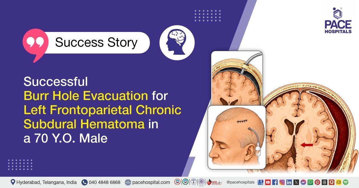
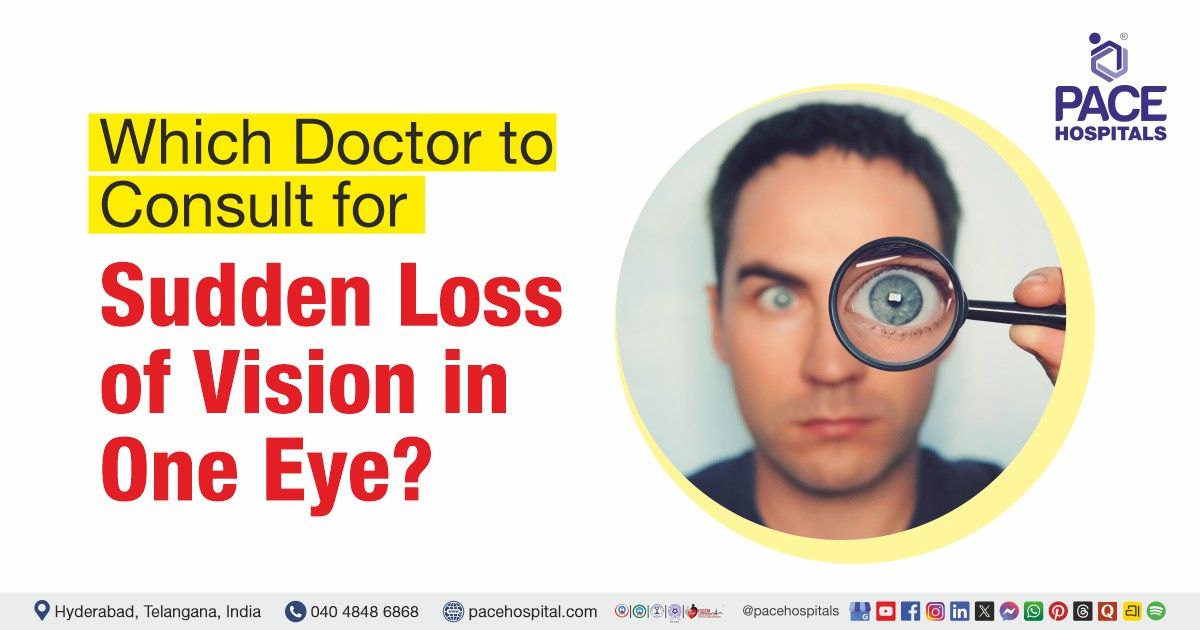
Which Doctor to Consult for Sudden Loss of Vision in One Eye?
PACE Hospitals
Written by: Editorial Team
Medically reviewed by: Dr. S Pramod Kumar - Consultant Neurophysician & Neuromuscular Specialist
Introduction
Sudden loss of vision in one eye should never be ignored or left to improve on its own. It is a medical emergency that can signal a serious condition affecting the eye, optic nerve, brain, or blood vessels — and every minute matters.
Whether the vision loss affects part or all of your sight, is painful or painless, or happens along with symptoms such as a curtain-like shadow, flashes of light, floaters, severe headache, or other neurological signs, it needs urgent medical attention. Getting evaluated by a healthcare professional as soon as possible is important to identify the cause and prevent potential complications. Home remedies, over-the-counter eye drops, or delaying medical care for several days can increase the risk of permanent vision loss and other serious complications.
This article helps patients and families in Hyderabad and across India understand exactly which doctor to consult for sudden vision loss in one eye, what the common causes are, what tests and treatments are involved, and when to head straight to an emergency department. PACE Hospitals, Hyderabad provides 24/7 emergency eye care, with experienced Ophthalmologists, Retina Specialists, and
Neurologists ready to evaluate and manage these urgent cases.
Quick Answer
Sudden loss of vision in one eye is a medical emergency. Seek immediate care at an Emergency Department or urgent eye care center and consult an Ophthalmologist without delay. A Retina Specialist may be required if there is suspicion of retinal detachment, bleeding inside the eye, blockage of retinal blood vessels, or diabetic eye disease. If vision loss occurs along with symptoms such as weakness, slurred speech, severe headache, confusion, or numbness, urgent
Neurology evaluation is also needed, as these may indicate a
stroke or other serious
neurological condition.
What Does Sudden Loss of Vision in One Eye Mean?
Sudden vision loss in one eye refers to any rapid, unexpected decrease or complete loss of sight in one eye — occurring over seconds, minutes, or hours. It may feel like a curtain dropping across the field of vision, a dark shadow covering part of the view, a sudden black or grey patch, blurring that does not clear, or a complete loss of light perception in one eye.
The loss may be:
- Partial — only part of the field of vision is affected
- Complete — total blindness in the affected eye
- Transient — vision returns after a few seconds or minutes but should never be ignored
- Painless — most causes of sudden vision loss are painless, which can falsely reassure patients
- Painful — some causes, such as acute angle-closure glaucoma, involve severe eye pain
Sudden vision loss in one eye is never caused by eye strain, screen use, or fatigue. It always requires urgent medical evaluation.
Why Sudden One-Eye Vision Loss Is an Emergency?
The eye is a direct extension of the brain. Blood vessels, nerves, and neural pathways connecting the eye to the brain are closely interconnected. A sudden loss of vision in one eye can be the result of a blockage in a retinal blood vessel (similar to a stroke), a detached retina, bleeding inside the eye, damage to the optic nerve, or an actual stroke or TIA (transient ischaemic attack) affecting the brain.
The American Academy of Ophthalmology states that sudden visual loss needs to be treated as a medical emergency until proven otherwise. The National Eye Institute (NIH) classifies retinal detachment as a medical emergency requiring immediate treatment to prevent permanent blindness. The American Stroke Association lists sudden vision loss as a core warning sign of stroke and TIA.
The window for effective treatment in many of these conditions is extremely narrow — for retinal artery occlusion (eye stroke), any intervention must ideally begin within 4 to 6 hours of onset. For retinal detachment, early surgical repair significantly improves the chance of preserving useful vision. Delayed evaluation can result in permanent, irreversible vision loss.
Do not wait. Do not drive yourself if you are experiencing active vision loss. Seek emergency care immediately.
Doctor Selection Guide
The table below provides a practical guide on which doctor to consult based on your situation.
| Situation | First Doctor to Consult | Specialist Needed If |
|---|---|---|
| Sudden, painless vision loss in one eye | Emergency Dept + Ophthalmologist (urgent) | Retina Specialist if retinal cause suspected |
| Curtain or shadow over vision | Emergency Ophthalmologist | Retina Specialist (retinal detachment, tear) |
| Flashes of light and new floaters | Urgent Ophthalmologist | Retina Specialist if tear or detachment found |
| Vision loss + severe headache | Emergency Department immediately | Neurologist (stroke, raised intracranial pressure, GCA) |
| Vision loss + weakness, slurred speech, facial droop | Emergency Dept (FAST protocol) | Neurologist / Stroke specialist |
| Vision loss + severe eye pain, red eye, nausea | Emergency Ophthalmologist | Ophthalmologist (acute angle-closure glaucoma) |
| Vision loss in a diabetic patient | Emergency Ophthalmologist | Retina Specialist (vitreous hemorrhage, diabetic retinopathy) |
| Vision loss + jaw pain, scalp tenderness in elderly | Emergency Department | Rheumatologist + Ophthalmologist (giant cell arteritis) |
| Vision loss after eye or head injury | Emergency Department | Ophthalmologist + Trauma team |
| Transient vision loss (returns within minutes) | Emergency Department immediately | Neurologist + Cardiologist (TIA, heart rhythm problem) |
| Vision loss + high blood pressure | Emergency Department | Internal Medicine + Ophthalmologist |
| Vision loss in young adult with eye pain | OphthalmologistOphthalmologist | Neurologist (optic neuritis, MS workup) |
When to Go to Emergency Immediately?
Go to an Emergency Department without delay if you experience any of the following:
- Sudden, complete or near-complete loss of vision in one eye
- Curtain-like shadow or black veil descending over your vision
- Sudden vision loss with weakness, numbness, or paralysis of the face, arm, or leg
- Vision loss with difficulty speaking, understanding speech, or slurred words
- Vision loss with severe headache — especially the worst headache of your life
- Sudden vision loss with facial drooping or loss of balance
- Transient (temporary) loss of vision that resolved — a possible TIA warning sign
- Vision loss after a direct injury to the eye or head
- Vision loss with jaw pain or scalp tenderness in a person over 50 years of age
Do not wait for symptoms to settle. Do not attempt home treatments. Time is critical.
When to See an Ophthalmologist?
An Ophthalmologist (eye specialist / eye surgeon) is the primary specialist to consult for any sudden loss of vision in one eye. An Ophthalmologist examines all structures of the eye — the cornea, lens, vitreous, retina, optic nerve, and eye pressure — using specialized instruments including a slit lamp, dilated fundus examination, OCT, and fluorescein angiography.
Consult an Ophthalmologist urgently for:
- Any sudden change in vision in one eye, even if partial
- Flashes of light or sudden increase in floaters
- A curtain or shadow over part of the visual field
- Red eye with pain and sudden vision blur (may indicate acute glaucoma)
- Sudden vision loss in a known diabetic patient
- Eye pain with or without vision change
- Vision loss following any eye injury
- Sudden decrease in vision in the elderly (to rule out giant cell arteritis)
An Ophthalmologist will perform a thorough dilated eye examination and determine whether a Retina Specialist, Neurologist, or other specialist consultation is needed.
When to See a Retina Specialist?
A Retina Specialist is an Ophthalmologist with advanced subspecialty training in conditions affecting the retina, vitreous, and the back portion of the eye. You may need to see a Retina Specialist if your Ophthalmologist finds or strongly suspects any of the following retinal causes of sudden vision loss:
- Retinal Detachment
- Retinal Tear — a small break that may lead to detachment if untreated
- Central Retinal Artery Occlusion (CRAO) — blockage of the main retinal artery; often called an "eye stroke"
- Branch Retinal Artery Occlusion (BRAO) — blockage causing partial field loss
- Central Retinal Vein Occlusion (CRVO) — blockage of the main vein draining the retina
- Branch Retinal Vein Occlusion (BRVO)
- Vitreous Hemorrhage — bleeding into the vitreous, common in diabetic patients
- Diabetic macular edema or diabetic retinopathy with sudden deterioration
- Macular hole or epiretinal membrane with sudden central vision change
According to the American Society of Retina Specialists (ASRS), retinal artery occlusion presents as sudden painless vision loss and must be referred promptly to an emergency room for stroke risk evaluation.
When to See a Neurologist?
A Neurologist may be required when the cause of sudden vision loss is related to the optic nerve, the brain's visual pathways, or the blood vessels supplying the brain. Consult a Neurologist — often through the Emergency Department — for:
- Vision loss with weakness, numbness, paralysis — possible stroke or TIA
- Vision loss with facial drooping, slurred speech, or confusion
- Vision loss with severe headache (possible brain bleed or raised intracranial pressure)
- Transient vision loss in one eye that fully resolved — possible amaurosis fugax (TIA warning sign)
- Optic neuritis — inflammation of the optic nerve with pain on eye movement; may be an early sign of multiple sclerosis
- Anterior Ischemic Optic Neuropathy (AION) — reduced blood flow to the optic nerve head causing sudden partial vision loss
- Posterior vision pathway lesions causing visual field defects
The American Stroke Association recognizes sudden vision loss as a core stroke warning symptom. If stroke is suspected, the patient must be transported to an Emergency Department immediately for CT or MRI brain imaging and urgent stroke team evaluation.
When Internal Medicine or Cardiology May Be Involved?
Many causes of sudden vision loss in one eye are driven by systemic disease. After emergency eye and neurological care, an Internal Medicine or Cardiology specialist may be involved in identifying and managing the underlying cause.
Systemic conditions that may cause sudden vision loss include:
- Diabetes mellitus — causes diabetic retinopathy, vitreous hemorrhage, and retinal vessel occlusion
- Hypertension (high blood pressure) — hypertensive retinopathy, retinal vessel occlusion, hypertensive crisis
- High cholesterol / hyperlipidaemia — cholesterol emboli can block retinal arteries
- Atrial fibrillation — blood clots from the heart may travel to the retinal artery or brain
- Carotid artery disease — narrowing reduces blood supply to the eye and brain
- Blood clotting disorders — increase the risk of vascular occlusion in the eye
- Giant cell arteritis / autoimmune vasculitis — requires emergency steroid treatment
Cardiologists may perform echocardiography and cardiac monitoring to detect embolic sources. Internal Medicine physicians manage the underlying systemic conditions that put patients at continued risk.
Sudden Vision Loss with Flashes and Floaters
Flashes and floaters are important warning signs that the retina may be at risk. Flashes of light occur when the vitreous gel pulls on the retina. New floaters — especially a sudden shower of dark spots, strings, or a web-like pattern — may indicate vitreous hemorrhage, a retinal tear, or the early stage of retinal detachment.
According to the National Eye Institute, a sudden increase in floaters or flashes in one eye is a medical emergency requiring immediate evaluation. Any sudden new onset of flashes and floaters — especially accompanied by a shadow or reduced vision — must be evaluated the same day.
Do not assume flashes and floaters are benign. They require urgent same-day evaluation by an Ophthalmologist or Emergency Department.
Curtain-Like Shadow over Vision
A curtain-like shadow or dark veil descending from the top, side, or bottom of the visual field is one of the most classic symptoms of retinal detachment. The detached portion of the retina can no longer process light, causing a corresponding dark area in the patient's field of vision.
A dark "curtain" or shadow in vision is an urgent warning sign requiring immediate evaluation. A curtain-like shadow may also occur in amaurosis fugax — a transient episode where a shade seems to cover one eye for seconds to minutes, which is a warning sign of impending stroke or TIA.
In either case — retinal detachment or amaurosis fugax — go to the Emergency Department immediately without delay.
Sudden Vision Loss with Eye Pain
Sudden vision loss accompanied by severe eye pain, redness, nausea, or vomiting suggests acute angle-closure glaucoma — a true ocular emergency. In this condition, the drainage angle of the eye becomes suddenly blocked, causing rapid and dangerous elevation of intraocular pressure (IOP) that can cause permanent optic nerve damage and blindness within hours if untreated.
Acute angle-closure glaucoma usually develops suddenly and can cause severe pain in one eye or on one side of the head, blurred vision, rainbow-colored halos around lights, nausea, and vomiting. It is a medical emergency that requires immediate treatment, as vision loss can occur within a short period if the condition is not treated promptly.
Sudden vision loss with severe eye pain is a medical emergency. Go to the Emergency Department or a 24-hour eye emergency service immediately.
Sudden Vision Loss without Pain
Many of the most serious causes of sudden vision loss in one eye are completely painless. The absence of pain does not mean the cause is minor or that waiting is acceptable.
Painless sudden vision loss causes include:
- Central Retinal Artery Occlusion (CRAO) — sudden, severe, painless vision loss
- Retinal Vein Occlusion — gradual or sudden painless blurring
- Vitreous Hemorrhage — painless blurring or floaters
- Retinal Detachment — painless curtain or shadow over vision (flashes and floaters may precede it)
- Anterior Ischemic Optic Neuropathy (AION) — painless sudden reduction of vision, often in one quadrant
- Giant Cell Arteritis — sudden painless vision loss in elderly patients (though headache and jaw pain may be present)
Painless sudden vision loss must be treated with the same urgency as painful vision loss. Seek emergency evaluation immediately.
Sudden Vision Loss in Diabetes
People living with diabetes mellitus are at significantly higher risk for several eye conditions that can cause sudden vision loss. According to the National Eye Institute, diabetic retinopathy can cause vision loss and blindness and affects blood vessels in the retina.
In a diabetic patient, sudden vision loss may be due to:
- Vitreous Hemorrhage — bleeding from fragile new blood vessels (neovascularization) into the vitreous cavity
- Tractional Retinal Detachment — scar tissue pulls the retina away from the back of the eye
- Diabetic Macular Edema — swelling at the center of the retina causing blurring
- Retinal Artery or Vein Occlusion — diabetes increases the risk of clot formation in retinal vessels
Any diabetic patient who experiences sudden vision change in one eye must seek emergency ophthalmology evaluation the same day. A Retina Specialist will likely be involved. Do not delay evaluation assuming it is a blood sugar fluctuation.
Sudden Vision Loss with Headache
The combination of sudden vision loss and headache should raise immediate concern for several serious conditions:
- Giant Cell Arteritis (GCA) / Temporal Arteritis — affects patients over 50; symptoms include new-onset severe headache (especially at the temples), scalp tenderness, jaw pain on chewing, and sudden vision loss. Approximately 20–30% of GCA patients experience visual disturbances. Untreated GCA can cause permanent blindness — often in both eyes. Emergency treatment with high-dose steroids is required.
- Stroke or Subarachnoid Hemorrhage — a sudden thunderclap headache described as the worst of one's life, combined with vision changes, is a red-flag emergency. Call emergency services immediately.
- Hypertensive Crisis — severely elevated blood pressure can cause hypertensive retinopathy, optic disc edema, and vision loss along with severe headache.
- Raised Intracranial Pressure — from various causes, leading to visual changes and headache.
Any vision loss combined with a new or unusually severe headache requires emergency evaluation without delay.
Eye Stroke and Retinal Vessel Blockage
An "eye stroke" is the common term for Central Retinal Artery Occlusion (CRAO) — a blockage of the main artery supplying blood and oxygen to the retina. It is the ocular equivalent of a brain stroke.
According to the American Society of Retina Specialists, CRAO results in sudden, painless, severe vision loss in one eye. The blockage is typically caused by a blood clot (thrombus) or a cholesterol embolus. The same risk factors that cause heart attacks and strokes — hypertension, diabetes, high cholesterol, atrial fibrillation, and carotid artery disease — also cause CRAO.
CRAO is associated with an elevated short-term risk of stroke. The ASRS recommends that any patient with CRAO or BRAO be referred promptly to the emergency room to evaluate for stroke risk.
Retinal Vein Occlusion (RVO) occurs when the vein draining the retina is blocked, causing blood and fluid to leak into the retina and leading to sudden blurring or vision loss. Both CRVO and BRVO are associated with high blood pressure, diabetes, and high cholesterol.
Treatment for retinal vessel occlusions may include anti-VEGF injections into the eye, laser treatment, and management of underlying vascular risk factors.
Retinal Detachment Warning Signs
Retinal detachment occurs when the retina separates from the back wall of the eye, depriving it of blood supply and causing vision loss. It is a surgical emergency.
According to the National Eye Institute, symptoms of retinal detachment include:
- A sudden large increase in floaters (dark spots, strings, or a shower of specks)
- Flashes of light in one eye
- A dark shadow or "curtain" on the sides or in the middle of the visual field
Risk factors for retinal detachment include:
- Severe short-sightedness (high myopia)
- Previous eye surgery or eye injury
- Family history of retinal detachment
- Prior retinal tear or detachment in the other eye
- Posterior Vitreous Detachment (PVD) — especially if complicated by a retinal tear
Treatment involves surgical reattachment through vitrectomy, scleral buckling, pneumatic retinopexy, laser, or cryotherapy, depending on the type and extent of detachment.
Causes and Conditions: Quick Reference Table
| Condition / Cause | Common Features | Doctor / Specialist | Why? |
|---|---|---|---|
| Central Retinal Artery Occlusion (CRAO) | Sudden, painless, severe vision loss; cherry-red spot on exam | Emergency Dept + Ophthalmologist + Retina Specialist | Eye stroke; 4–6 hour treatment window; stroke risk evaluation needed |
| Branch Retinal Artery Occlusion (BRAO) | Loss of part of visual field, usually painless | Emergency Dept + Ophthalmologist + Retina Specialist | Stroke risk evaluation; treat urgently |
| Central Retinal Vein Occlusion (CRVO) | Sudden painless blurring; dilated, tortuous retinal veins | Ophthalmologist + Retina Specialist | Anti-VEGF or laser treatment; manage vascular risk factors |
| Retinal Detachment | Curtain-like shadow, flashes, floaters, vision loss | Emergency Ophthalmologist + Retina Specialist | Surgical emergency |
| Retinal Tear | Flashes and floaters without shadow yet | Urgent Ophthalmologist + Retina Specialist | Laser before detachment develops |
| Vitreous Hemorrhage | Sudden dark floaters, blurring; often in diabetics | Ophthalmologist + Retina Specialist | Retinal cause must be ruled out; vitrectomy if needed |
| Acute Angle-Closure Glaucoma | Severe eye pain, red eye, nausea, rainbow halos, sudden vision blur | Emergency Ophthalmologist | IOP must be lowered urgently to prevent optic nerve damage |
| Optic Neuritis | Sudden vision loss, pain on eye movement, young adults | Ophthalmologist + Neurologist | MS workup; steroid treatment if indicated |
| Anterior Ischemic Optic Neuropathy (AION) | Painless, sudden vision loss (upper or lower field), swollen optic disc | Ophthalmologist | Arteritic (GCA) vs. non-arteritic; urgent steroids if GCA |
| Giant Cell Arteritis (GCA) | Elderly; headache, jaw pain, scalp tenderness, vision loss | Emergency Dept + Ophthalmologist + Rheumatologist | High-dose IV steroids urgently; prevent bilateral blindness |
| Amaurosis Fugax (TIA of the eye) | Transient monocular vision loss that resolves; curtain-like | Emergency Dept + Neurologist + Cardiologist | Stroke warning; urgent carotid imaging and cardiac workup |
| Stroke / TIA | Vision loss + weakness, speech difficulty, facial droop, confusion | Emergency Dept + Neurologist (Stroke team) | FAST protocol; thrombolysis window |
| Diabetic Vitreous Hemorrhage | Sudden blurring or dark floaters in diabetic patient | Emergency Ophthalmologist + Retina Specialist | Vitrectomy may be needed; control underlying diabetes |
| Hypertensive Retinopathy | Vision changes with very high BP, headache | Emergency Dept + Internal Medicine + Ophthalmologist | BP control; prevent optic nerve and retinal damage |
Red-Flag Symptoms Checklist
The following symptoms require immediate emergency evaluation — do not delay:
- Sudden complete or partial loss of vision in one eye
- Curtain-like shadow or dark veil descending over the field of vision
- Sudden, large increase in floaters or a shower of dark spots
- Flashes of light in one eye, especially if new or persistent
- Sudden vision loss with no pain (many serious causes are painless)
- Severe eye pain with vision loss, redness, and nausea
- Vision loss combined with severe headache
- Vision loss combined with weakness, numbness, or paralysis
- Vision loss combined with facial drooping or slurred speech
- Vision loss combined with confusion or loss of balance
- Transient vision loss that returned — even if the episode lasted only seconds
- Vision loss in a diabetic patient or hypertensive patient
- Jaw pain on chewing, scalp tenderness, or new headache in a patient over 50
- Vision loss following a direct eye or head injury
Any one of these symptoms is sufficient reason to go to the Emergency Department immediately.
Tests Doctors May Recommend
Eye Examination Tests
- Dilated Fundus Examination — the most important initial test; examines retina, optic nerve, and retinal vessels
- Slit Lamp Examination — detailed examination of the front and back of the eye
- Intraocular Pressure (IOP) Measurement (Tonometry) — critical for diagnosing acute angle-closure glaucoma
- Visual Acuity Testing — measures the clarity of vision and severity of vision loss
- Visual Field Testing (Perimetry) — maps areas of vision loss to localize the problem
- Optical Coherence Tomography (OCT) — non-invasive cross-sectional scan of the retina and optic nerve
- Fluorescein Angiography (FFA) — assesses retinal blood flow; used for retinal vessel occlusions
- B-Scan Ultrasound — used when the view into the eye is blocked to assess for retinal detachment
Blood Tests
- ESR and C-Reactive Protein (CRP) — mandatory in all patients over 50 with sudden vision loss; elevated in giant cell arteritis
- Complete Blood Count (CBC) — checks for blood disorders
- Blood Glucose and HbA1c — to assess diabetes control
- Lipid Profile — elevated cholesterol is a risk factor for retinal artery occlusion
- Coagulation Profile / Clotting Studies — to rule out blood clotting disorders
Neuroimaging
- MRI Brain (with diffusion-weighted imaging) — most sensitive test for acute stroke, demyelinating lesions (MS/optic neuritis)
- CT Scan Brain — rapid initial test to rule out brain bleeding
- MR Angiography or CT Angiography — images blood vessels in the brain and neck
Cardiovascular Tests
- Carotid Ultrasound (Doppler) — detects narrowing of the carotid arteries
- ECG — detects atrial fibrillation and heart rhythm disorders
- Echocardiogram — detects clots or structural heart problems as embolic sources
- Holter Monitor — detects intermittent arrhythmias over 24 hours
Temporal Artery Biopsy
Performed when giant cell arteritis is suspected. A small segment of the temporal artery is removed and examined under a microscope. This remains the gold standard for confirming GCA.
Treatment Options
Treatment depends entirely on the underlying cause of sudden vision loss. Prompt evaluation is the first and most important step — no home treatment can substitute for professional medical assessment.
Retinal Detachment
- Laser photocoagulation (laser retinopexy) — for retinal tears without detachment
- Cryopexy (freezing treatment) — seals retinal tears
- Pneumatic retinopexy — gas bubble injected to push the retina back into place
- Scleral buckling — a band placed around the eye to support the detached retina
- Vitrectomy — removal and replacement of the vitreous gel; used for complex detachments and vitreous hemorrhage
Retinal Artery and Vein Occlusion
- Anti-VEGF intravitreal injections— main treatment for retinal vein occlusion with macular edema
- Intravitreal steroid injections — alternative for inflammation-related edema
- Laser treatment (panretinal photocoagulation) — for severe or proliferative complications
- Management of underlying vascular risk factors — blood pressure, cholesterol, diabetes, cardiac arrhythmias
Acute Angle-Closure Glaucoma
- Immediate IOP reduction with topical and systemic medications
- Laser peripheral iridotomy (LPI) — creates a small opening in the iris to restore fluid drainage
- Surgical intervention if laser treatment is insufficient
Optic Neuritis
- High-dose intravenous corticosteroids may speed visual recovery
- Neurological evaluation and MRI for multiple sclerosis workup
Giant Cell Arteritis
- Emergency initiation of high-dose systemic corticosteroids
- Long-term tapering steroid regimen with monitoring of ESR and CRP
- Temporal artery biopsy for definitive diagnosis
Vitreous Hemorrhage
- Observation in mild cases — blood may clear spontaneously
- Vitrectomy surgery to remove blood-filled vitreous if clearance is delayed or retinal detachment is present
- Treatment of underlying diabetic retinopathy with anti-VEGF injections and laser
Stroke / TIA
- Emergency CT or MRI brain imaging
- Thrombolytic therapy (clot-busting medication) if within the treatment window for ischaemic stroke
- Antiplatelet therapy for TIA
- Anticoagulation if atrial fibrillation is identified
- Stroke unit care, rehabilitation, and secondary prevention
Eye and Emergency Specialists at PACE Hospitals, Hyderabad
PACE Hospitals, Hyderabad, offers comprehensive emergency and specialist eye care for patients with sudden vision loss in one eye. The hospital is equipped with advanced diagnostic facilities and experienced specialists managing urgent and complex ophthalmic and neurological emergencies.
Specialists involved in sudden vision loss care at PACE Hospitals include:
- Ophthalmologists — for comprehensive emergency eye evaluation and management of retinal, glaucoma, and optic nerve conditions
- Retina Specialists (Vitreo-Retinal Surgeons) — for retinal detachment surgery, vitrectomy, anti-VEGF injections, laser treatment, and management of retinal vessel occlusions and vitreous hemorrhage
- Neurologists — for evaluation of stroke, TIA, optic neuritis, and other neurological causes of sudden vision loss
- Interventional Cardiologists and Internal Medicine Specialists — for management of atrial fibrillation, carotid artery disease, hypertension, and diabetes
- Emergency Medicine Specialists — available 24/7 for immediate assessment and stabilization
PACE Hospitals provides 24-hour emergency services with advanced imaging including MRI, CT angiography, OCT, fluorescein angiography, and B-scan ultrasound to support rapid diagnosis and treatment.
Why Choose PACE Hospitals?
- 24/7 Emergency Department with immediate ophthalmology and neurology response
- Advanced Vitreo-Retinal Surgery facilities for retinal detachment, vitrectomy, and complex retinal conditions
- Comprehensive Eye Care including dilated fundus examination, OCT, fluorescein angiography, and B-scan ultrasound
- State-of-the-art neuroimaging — MRI with DWI, CT, CT angiography, and MR angiography
- Multidisciplinary Stroke Team for integrated stroke and TIA management
- Cardiology and Internal Medicine specialists for systemic causes of vision loss
- Patient-friendly care with multilingual teams, clear communication, and streamlined emergency pathways
- Convenient location in Hyderabad with easy accessibility for patients across Telangana and Andhra Pradesh
Key Takeaway
- Sudden loss of vision in one eye is a medical emergency — not a symptom to watch at home.
- The first and most important step is to go to an Emergency Department or urgent eye care service immediately.
- An Ophthalmologist is the primary specialist to consult.
- A Retina Specialist is needed for retinal detachment, retinal vessel occlusion, vitreous hemorrhage, or diabetic eye emergencies.
- A Neurologist must be involved if vision loss is associated with stroke symptoms — weakness, slurred speech, facial drooping, severe headache, or confusion.
- Internal Medicine and Cardiology are involved when diabetes, hypertension, high cholesterol, or heart rhythm disorders are the underlying drivers.
- Time is critical — for some conditions, the treatment window is as short as 4 to 6 hours.
- Never assume painless vision loss is minor — many of the most serious causes are completely painless.
- Do not self-medicate or wait for vision to settle. Seek emergency care immediately.
Frequently Asked Questions (FAQs)
Which doctor should I consult for sudden vision loss in one eye?
Sudden vision loss in one eye requires immediate emergency evaluation. The first step is to go to an Emergency Department or a 24-hour eye emergency service. An Ophthalmologist is the primary specialist who will examine your eye urgently. Depending on the cause, you may be referred to a Retina Specialist (for retinal causes such as detachment, artery or vein blockage, or diabetic eye bleeding), a Neurologist (for stroke, optic neuritis, or TIA), or an Internal Medicine specialist (for diabetes or high blood pressure). At PACE Hospitals, Hyderabad, all these specialists are available 24/7 for emergency evaluation.
Is sudden loss of vision in one eye an emergency?
Yes, absolutely. Sudden loss of vision in one eye is a medical emergency. It can be caused by conditions such as retinal detachment, retinal artery occlusion (eye stroke), stroke or TIA, acute angle-closure glaucoma, or giant cell arteritis — all of which require urgent medical treatment. According to the National Eye Institute, retinal detachment is a medical emergency. The American Stroke Association identifies sudden vision loss as a stroke warning sign. Do not wait, do not observe at home — go to an Emergency Department immediately.
Can retinal detachment cause sudden vision loss?
Yes, retinal detachment is one of the most important causes of sudden vision loss in one eye, and it is a surgical emergency. The retina separates from the back wall of the eye, causing a dark curtain or shadow over the field of vision, often preceded by flashes of light and a sudden increase in floaters. According to the National Eye Institute, if retinal detachment is not treated promptly, more of the retina can detach, increasing the risk of permanent blindness. Surgery to re-attach the retina can preserve or restore vision if performed early.
What does a curtain over vision mean?
A curtain-like shadow or dark veil descending over part of the field of vision is a classic warning sign of retinal detachment. It occurs because the detached part of the retina is no longer functioning. This is a surgical emergency requiring immediate evaluation by an Ophthalmologist and likely a Retina Specialist. A similar curtain-like vision loss can also occur in amaurosis fugax — a transient episode caused by brief interruption of blood supply to the eye — which is a warning sign of an impending stroke or TIA. In either case, go to the Emergency Department immediately.
What tests are done for sudden vision loss?
The evaluation of sudden vision loss involves several urgent tests to find the cause. An Ophthalmologist may first perform a dilated fundus examination, slit lamp examination, and intraocular pressure measurement. OCT provides detailed images of the retina, while fluorescein angiography is used to assess blood flow in the retinal vessels. If the inside of the eye cannot be clearly seen, a B-scan ultrasound may be used. Blood tests may include ESR, CRP, blood glucose, lipid profile, and clotting studies. When a stroke or other neurological condition is suspected, investigations such as MRI brain (with DWI), CT brain, and carotid ultrasound may be recommended. ECG and echocardiography can help identify cardiac sources of emboli. These diagnostic tests and specialist evaluations are available at PACE Hospitals, Hyderabad.
What should I avoid if I suddenly lose vision?
If you suddenly lose vision in one eye, do not wait at home hoping the vision will return. Do not apply eye drops, rub the eye, or attempt any home remedies. Do not assume the vision loss is due to eye strain, screen use, or fatigue. Do not drive yourself to the hospital if your vision is affected — ask a family member or call for emergency transport. Do not delay seeking care even if the vision partially returns, as transient vision loss is a warning sign that must be evaluated urgently. Do not take any medications without medical advice. Go to the Emergency Department immediately.
Which is the best hospital for sudden vision loss in Hyderabad?
PACE Hospitals, Hyderabad is a leading multi-specialty hospital providing 24/7 emergency eye care, retina surgery, and neurology services for patients with sudden vision loss in one eye. The hospital offers advanced diagnostic facilities including OCT, fluorescein angiography, B-scan ultrasound, MRI, and CT angiography, along with experienced Ophthalmologists, Retina Specialists, Neurologists, Cardiologists, and Emergency Medicine specialists. PACE Hospitals is equipped to handle the full spectrum of eye emergencies — from retinal detachment and eye stroke to acute glaucoma and neurological vision loss — under one roof, ensuring rapid, coordinated, and comprehensive care.
Should I see an ophthalmologist for sudden vision loss?
Yes. An Ophthalmologist is the primary specialist for sudden vision loss in one eye. They will perform a dilated eye examination, measure intraocular pressure, assess the retina and optic nerve, and determine the cause of vision loss. If a retinal cause is found (detachment, vitreous hemorrhage, or vessel blockage), they may refer you to a Retina Specialist. If a neurological cause is suspected (stroke, optic neuritis), they will refer you to a Neurologist. Always seek urgent Ophthalmology evaluation — do not wait for a routine appointment.
When should I see a retina specialist?
You may need to see a Retina Specialist if your Ophthalmologist suspects that the sudden vision loss is coming from a problem in the retina, the light-sensitive layer at the back of the eye. Conditions that often require urgent retinal evaluation include retinal detachment, retinal tears, retinal artery or vein occlusion (sometimes called an eye stroke), vitreous hemorrhage, and complications related to diabetic retinopathy. Retina Specialists use advanced tests such as OCT, fluorescein angiography, and B-scan ultrasound to diagnose these conditions and can perform specialized retinal procedures and surgeries when needed. Symptoms such as a curtain-like shadow over your vision, a sudden increase in floaters, flashes of light, or sudden blurred vision—especially in people with diabetes—should be evaluated by a Retina Specialist as soon as possible.
Can diabetes cause sudden vision loss?
Yes. Diabetes can increase the risk of sudden vision loss in one eye for several reasons. In diabetic retinopathy, fragile abnormal blood vessels can bleed into the vitreous cavity, leading to sudden blurry vision, dark spots, or floaters. In more advanced cases, scar tissue may form and pull on the retina, causing a tractional retinal detachment. Diabetic macular edema can also affect the central part of vision and lead to noticeable blurring. In addition, people with diabetes have a higher risk of retinal artery and retinal vein occlusions. Any sudden change in vision in a person with diabetes should be treated as an emergency and evaluated by an Ophthalmologist as soon as possible.
Can high blood pressure cause sudden vision loss?
Yes. Severely elevated blood pressure can cause sudden vision loss through several pathways. Hypertensive retinopathy — damage to the retinal blood vessels — can cause retinal hemorrhages, cotton wool spots, and macular edema. A hypertensive crisis can lead to optic disc edema (swelling of the optic nerve head) and significant vision loss. High blood pressure is also a major risk factor for retinal artery occlusion (eye stroke) and retinal vein occlusion. In patients over 50, sudden vision loss with high blood pressure also raises suspicion for giant cell arteritis. Emergency evaluation is mandatory.
Can stroke cause vision loss in one eye?
Yes, stroke can cause sudden vision loss in one eye. When a stroke or TIA affects blood supply to the eye through the ophthalmic or central retinal artery, it causes sudden, often painless loss of vision. When the vision loss is transient (returning after seconds to minutes) it is called amaurosis fugax or a TIA of the eye — a serious warning sign that a full stroke may follow. The American Stroke Association lists sudden vision loss as a core stroke warning symptom. If vision loss occurs with weakness, numbness, slurred speech, facial droop, or severe headache, call emergency services immediately — use the FAST protocol.
Is OCT needed for sudden vision loss?
Yes, OCT (Optical Coherence Tomography) is one of the key tests used to evaluate sudden vision loss in one eye. It is a non-invasive imaging technique that provides detailed cross-sectional images of the retina, macula, and optic nerve. OCT can help identify conditions such as retinal vein occlusion with macular edema, diabetic macular edema, optic nerve swelling seen in optic neuritis or anterior ischemic optic neuropathy, retinal detachment, vitreous hemorrhage, and macular holes. The information obtained from OCT helps doctors determine the cause of vision loss and plan appropriate treatment, including the need for anti-VEGF injections. OCT is available at PACE Hospitals, Hyderabad, as part of the comprehensive eye evaluation process.
Is MRI needed for vision loss?
MRI of the brain is recommended when a neurological cause of sudden vision loss is suspected. MRI with diffusion-weighted imaging (DWI) is the most sensitive test for detecting acute brain stroke — including strokes affecting the visual pathway. It also detects demyelinating lesions associated with optic neuritis and multiple sclerosis, brain tumors causing visual field defects, and vascular abnormalities. MR Angiography images blood vessels of the brain and neck. CT scan is usually performed first in emergency settings to rule out brain bleeding. MRI is available 24/7 at PACE Hospitals for urgent neurological assessment.
Can sudden vision loss be treated?
Whether sudden vision loss can be treated depends on the underlying cause and how quickly treatment is started. In cases of retinal detachment, prompt surgery can often preserve or even restore vision. For retinal vein occlusion, anti-VEGF injections may help reduce macular edema and improve vision outcomes. In giant cell arteritis, urgent high-dose corticosteroids are required to prevent further vision loss and protect the other eye. Acute angle-closure glaucoma needs immediate pressure-lowering treatment to avoid permanent damage. If a stroke is responsible, thrombolysis may be possible if the patient reaches the hospital within the treatment window. In all cases, time is critical—the earlier treatment begins, the better the chance of saving vision.
Conclusion
Sudden loss of vision in one eye is one of the most urgent symptoms a person can experience. Whether it presents as a dark curtain descending over the field of vision, a sudden shower of floaters, painless total blackout, brief transient blindness, or vision loss combined with headache and neurological signs — every scenario demands immediate emergency medical evaluation.
The primary doctor to consult is an Ophthalmologist — ideally through an Emergency Department or 24-hour eye care service. Depending on the underlying cause, a Retina Specialist will be involved for retinal conditions, a Neurologist for stroke and optic nerve disease, and Internal Medicine or Cardiology for systemic vascular conditions.
This is not a condition where a scheduled appointment in a few days is appropriate. Time lost is vision lost — and in many cases, permanent vision loss can be prevented with prompt, expert care.
PACE Hospitals, Hyderabad provides the integrated, rapid, expert emergency eye care needed when it matters most. Do not delay — if you or someone you know experiences sudden vision loss, seek emergency care right away.
Share on
Request an appointment
Fill in the appointment form or call us instantly to book a confirmed appointment with our super specialist at 04048486868
Appointment request - health articles
Recent Articles