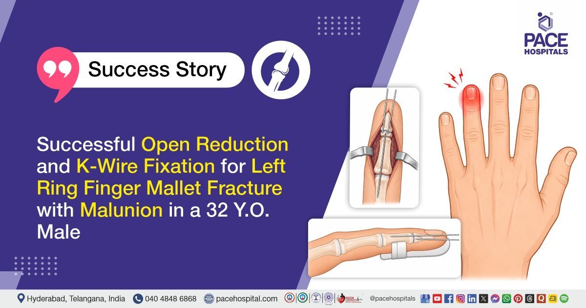
Successful Open Reduction and K-Wire Fixation for Left Ring Finger Mallet Fracture with Malunion
PACE Hospitals
PACE Hospitals' expert Orthopaedic team successfully performed an Open Reduction, Corticotomy, and Multiple K-wire Fixation with the extension block technique under C-arm guidance on a 32-year-old male patient diagnosed with an old mallet fracture of the left ring finger with malunion. The procedure was undertaken to correct the malunited fracture, restore proper alignment and stability of the distal phalanx, improve finger extension, and regain optimal hand function.
Chief Complaints
A 32-year-old male patient with a body mass index (BMI) of 22 presented to the Orthopaedic Department at PACE Hospitals, Hitech City, Hyderabad, with pain and deformity of the distal phalanx of the left ring finger, accompanied by restricted movement at the distal interphalangeal (DIP) joint. The patient had a history of blunt trauma following a fall on steps at home, which resulted in the injury.
Past Medical History
The patient had no significant past medical history and was not known to have any chronic illnesses such as diabetes mellitus, hypertension, cardiovascular disease, or other major systemic disorders. There was no history of previous surgeries, regular medication use, or known drug allergies. The patient's medical history was otherwise normal.
On Examination
On examination, the patient was conscious, coherent, and well-oriented, afebrile, and hemodynamically stable. Local examination of the left ring finger revealed deformity over the distal phalanx (the bone at the very tip of the finger) with associated swelling and tenderness. Active extension at the distal interphalangeal (DIP) joint was restricted, consistent with a mallet finger deformity. Distal neurovascular status was intact, with no evidence of sensory or vascular compromise. The remainder of the systemic examination was normal.
Diagnosis
Following the clinical evaluation, the Orthopaedic team at PACE Hospitals conducted a detailed assessment of the patient's complaints of pain and deformity over the distal phalanx of the left ring finger, associated with difficulty in movements of the distal interphalangeal (DIP) joint, following a history of blunt trauma due to a fall on steps at home.
A thorough clinical examination revealed deformity, swelling, and tenderness over the distal phalanx of the left ring finger, with inability to achieve complete active extension at the DIP joint. The distal neurovascular status was intact, and no significant abnormalities were noted on general physical examination.
Further clinical and radiological evaluation confirmed an old mallet fracture of the left ring finger with malunion. Based on the patient's clinical presentation, examination findings, and imaging results, the patient was diagnosed with an old mallet fracture of the left ring finger with malunion.
Based on these findings, the patient was advised to undergo
Left Ring Finger Old
Mallet Fracture Treatment in Hyderabad, India, with Malunion to correct the deformity, restore alignment and stability, improve DIP joint extension, relieve pain, restore finger function, and prevent long-term complications.
Medical Decision Making (MDM)
After a detailed evaluation by Dr. Anand Agroya, (Senior Orthopedic Consultant), the patient was assessed for pain and deformity over the distal phalanx of the left ring finger, associated with difficulty in extension at the distal interphalangeal (DIP) joint following a history of blunt trauma due to a fall on steps at home. Clinical examination revealed deformity, swelling, and tenderness over the affected region, with restricted active extension at the DIP joint. Distal neurovascular status was intact, and the patient was clinically stable with no significant systemic abnormalities. Radiological evaluation confirmed an old mallet fracture of the left ring finger with malunion.
Based on the clinical findings, imaging results, and functional impairment, it was determined that open reduction, corticotomy, and multiple K-wire fixation with the extension block technique under C-arm guidance was the most appropriate management approach. This decision was made to correct the malunion, restore anatomical alignment and stability of the distal phalanx, improve DIP joint extension, relieve pain, restore finger function, and prevent long-term deformity and functional impairment.
The patient and his family members were counselled regarding the diagnosis, the planned surgical procedure, fixation technique, potential risks and benefits, postoperative care, rehabilitation requirements, expected recovery course, and the importance of regular follow-up to achieve optimal functional outcomes.
Surgical Procedure
Following the diagnosis, the patient was scheduled to undergo Open Reduction, Corticotomy, and Multiple K-wire fixation in Hyderabad at PACE Hospitals with the extension block technique, under the supervision of the expert Orthopaedic Department.
The surgical procedure involved the following steps:
- Anaesthesia and Patient Preparation: The patient was taken to the operating room and placed under regional anaesthesia. The left hand was prepared and draped in a sterile manner. Proper positioning was ensured for optimal access to the left ring finger under C-arm guidance.
- Exposure and Identification: A surgical approach was made to the affected left ring finger. The malunited fracture site at the distal phalanx was carefully identified, and soft tissues were gently dissected to expose the fracture and deformity.
- Corticotomy and Fracture Mobilisation: A controlled corticotomy was performed to mobilise the malunited fragment. The fracture site was carefully manipulated to achieve proper alignment and correction of deformity under continuous C-arm imaging.
- Reduction and K-Wire Fixation: An open reduction was achieved, and the fracture fragments were accurately aligned. Stabilisation was performed using multiple K-wires, applied using the extension block technique to maintain proper alignment and restore distal phalanx stability.
- Final Assessment and Immobilisation: Final positioning and fixation were confirmed under C-arm guidance to ensure satisfactory reduction. The surgical site was irrigated and dressed in a sterile manner, and the finger was immobilised. The procedure was completed uneventfully.
Postoperative Care
Postoperatively, the patient was managed with intravenous medications to prevent infection, medications for pain relief, and supportive care to promote recovery. The operated hand was immobilised and elevated to reduce swelling and maintain fracture stability. Regular sterile dressing changes were performed to monitor wound healing and prevent complications. The patient remained hemodynamically stable and recovered well, following which he was discharged with advice for follow-up care.
Discharge Medications
Upon discharge, the patient was prescribed medications for prevention of infection at the surgical site for a short duration, medications for pain and inflammation control to reduce postoperative discomfort, and medications to protect the gastric lining and prevent irritation associated with treatment. These were advised for a limited period as part of routine postoperative care following surgical fixation of the finger fracture.
Advice on Discharge
The patient was advised to keep the surgical dressing dry and avoid allowing it to get wet.
Emergency Care
The patient was informed to contact the emergency ward at PACE Hospitals in case of any emergency or development of symptoms such as severe pain at the operated site, increasing swelling, fever, wound discharge, numbness, or discoloration of the left ring finger.
Review and Follow-up Notes
The patient was advised to return for follow-up with the Orthopaedic Doctor in Hyderabad at PACE Hospitals for dressing change after 2 days. He was also advised to undergo K-wire removal after 4 to 6 weeks post-surgery on an outpatient basis.
Conclusion
This case highlights an old mallet fracture of the left ring finger with malunion following blunt trauma. Management was done with open reduction, corticotomy, and multiple K-wire fixation using the extension block technique under C-arm guidance. The procedure aimed to restore anatomical alignment, improve distal interphalangeal joint function, and relieve pain. The postoperative course was uneventful, and the patient was discharged in a stable condition.
Surgical Management of Mallet Finger Deformity
Mallet finger is a condition characterized by loss of active extension at the distal interphalangeal (DIP) joint, leading to deformity and functional limitation of the finger. Management by an orthopaedic doctor / orthopaedic surgeon is aimed at restoring normal alignment of the extensor mechanism and improving finger function. Treatment options include operative methods that stabilize the distal phalanx and maintain proper joint positioning during healing, often with the assistance of imaging guidance to ensure accurate alignment and fixation. The primary objective of management is to restore DIP joint extension, correct deformity, and preserve hand function. In chronic or neglected cases, surgical correction may be required to address established deformity and improve joint mechanics, while appropriate immobilization and rehabilitation are essential to achieve optimal functional recovery and prevent long-term stiffness or recurrence.
Frequently Asked Questions (FAQs)
Is surgery needed for an old mallet fracture with malunion?
In an old mallet fracture with malunion, the broken bone fragment may heal in an improper position, causing finger deformity and difficulty in straightening the fingertip. When the deformity affects finger movement or daily hand use, surgery may be advised to realign the fracture and improve DIP joint function. In this case, surgery was done because the patient had pain, deformity and difficulty moving the left ring finger.
What does “open reduction, corticotomy and K-wire fixation” mean?
Open reduction means the surgeon opens the area and corrects the position of the malunited bone. Corticotomy means carefully cutting or loosening the healed bone area so it can be realigned properly. K-wire fixation means thin metal wires are used to hold the bone and joint in the corrected position until healing occurs.
Why was the extension block technique used in this mallet fracture?
The extension block technique is commonly used to hold the fingertip joint in a corrected extended position while the fracture fragment heals. It helps prevent the distal phalanx from bending again and gives stability to the repaired mallet fracture. This technique is useful when the fracture needs controlled reduction and temporary fixation. K-wires are commonly used to maintain this position during healing.
Why was C-arm guidance used during finger surgery?
C-arm guidance is a live X-ray imaging method used inside the operation theatre. It helps the surgeon check the bone alignment and K-wire position during the procedure. In small finger bones, accurate placement is very important because even a small change in alignment can affect fingertip movement and healing.
How long do K-wires usually stay after mallet fracture surgery?
K-wires are usually kept for around 4 to 6 weeks, depending on fracture healing and the surgeon’s assessment. Before removal, an X-ray may be advised to confirm that the bone has healed well. In this case, the discharge advice also mentions K-wire removal after 4 to 6 weeks on an outpatient basis.
Is K-wire removal painful or does it need another surgery?
K-wire removal is usually a small outpatient procedure and often does not require major surgery or hospital admission. The doctor removes the wires once the fracture has healed enough. Some patients may feel mild discomfort, but it is usually quick and manageable. After wire removal, finger movement exercises may be started as advised by the surgeon.
What precautions are important after mallet fracture K-wire fixation?
The dressing should be kept dry and clean, and the patient should avoid wetting the operated finger. The hand should be protected from accidental injury, pressure or pulling on the wires. Follow-up dressing and review visits are important to check wound healing and pin-site condition. Fever, increasing pain, swelling, discharge or redness around the wire site should be reported early.
When can the patient start moving the finger after this surgery?
Movement depends on the surgeon’s advice and the stability of fixation. Usually, nearby joints like the PIP and MCP joints may be moved earlier to reduce stiffness, while the DIP joint is protected until the K-wires are removed. After wire removal and confirmation of healing, gradual DIP joint exercises are started. Physiotherapy or guided exercises may be needed for better recovery.
Will the finger become completely normal after old mallet fracture correction?
The goal of surgery is to improve alignment, reduce deformity and restore better fingertip function. Since this was an old fracture with malunion, complete normal movement may take time and may not always be 100% like before injury. Some stiffness, mild extension lag or reduced flexibility can remain in a few patients. Regular follow-up and proper hand exercises help improve the final outcome.
What are the possible risks after mallet fracture surgery with K-wires?
Possible risks include stiffness, swelling, pin-site infection, delayed healing, wire loosening, residual deformity or reduced fingertip movement. These risks are usually reduced with proper dressing care, avoiding injury to the operated finger and attending scheduled follow-ups. K-wire fixation is a commonly used method for selected mallet fractures, but recovery depends on fracture age, alignment, healing response and rehabilitation.
Share on
Request an appointment
Fill in the appointment form or call us instantly to book a confirmed appointment with our super specialist at 04048486868
Appointment request - health articles
Recent Articles