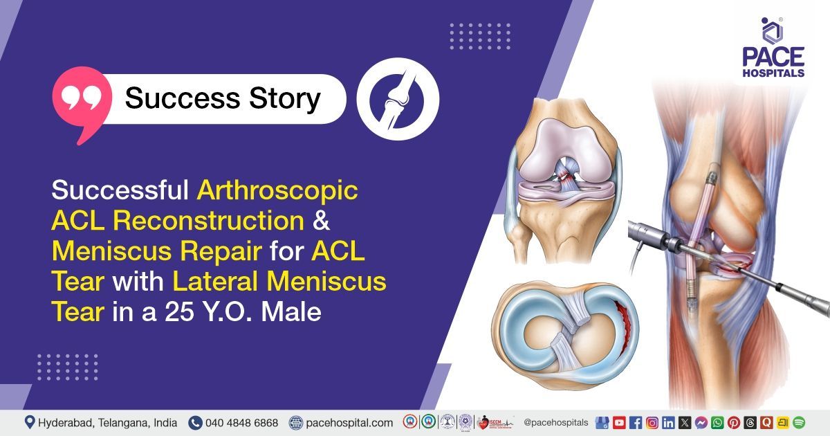
Successful Arthroscopic ACL Reconstruction & Meniscus Repair for ACL Tear with Lateral Meniscus Tear in a 25 Y.O. Male
PACE Hospitals
PACE Hospitals’ expert Orthopaedic team successfully performed a Lateral Meniscus Posterior Horn Radial Tear Repair followed by Right ACL Reconstruction using an Ipsilateral Hamstring (semitendinosus) Graft on a 25-year-old male patient. The patient was diagnosed with a complete mid-substance tear of the right anterior cruciate ligament, along with a complete radial tear of the posterior horn of the right lateral meniscus. The aim of the procedure was to restore knee stability, preserve meniscal integrity, reduce pain, and enable a safe return to daily activities and sports while minimizing the risk of long-term joint degeneration.
Chief Complaints
A 25-year-old male patient with a body mass index (BMI) of 22 presented to the Orthopaedic Department at PACE Hospitals, Hitech City, Hyderabad, with complaints of pain in the right knee, associated with difficulty in performing daily routine activities and a sense of instability, particularly while climbing stairs, following a history of self-fall from a bike.
Past Medical History
The patient was a known case of diabetes mellitus. There was no history of known drug allergies, and no other significant past medical or surgical history was reported.
On Examination
On examination, the patient presented with right knee joint tenderness, predominantly over the anterior joint line, along with marked restriction of range of motion due to pain. Special tests revealed a positive anterior drawer test, positive Lachman test, and positive pivot shift test, indicating anterior cruciate ligament insufficiency. The McMurray test was also positive, suggestive of associated meniscal injury.
There was no evidence of varus or valgus instability, and the posterior drawer test was negative. The patient demonstrated painful and restricted terminal flexion of the knee with associated functional instability. Generalized joint hyperextension was also noted involving multiple joints, including the knee, thumb, and elbow. Distal neurovascular status was intact with no deficits noted.
Diagnosis
Following the clinical evaluation, the Orthopaedics team at PACE Hospitals conducted a detailed assessment focusing on the patient’s complaints of pain in the right knee associated with difficulty in walking, performing daily routine activities, and a sense of instability, particularly while climbing stairs, following a history of self-fall from a bike.
A comprehensive clinical and systemic examination was carried out to determine the severity of the condition. Examination of the right knee revealed tenderness over the anterior joint line with gross restriction of range of motion due to pain. Special clinical tests, including anterior drawer test, Lachman test, and pivot shift test, were positive, indicating anterior cruciate ligament insufficiency. The McMurray test was also positive, suggestive of associated meniscal injury. There was no evidence of varus or valgus instability, and the posterior drawer test was negative. There was no distal neurovascular deficit in the affected limb.
Radiological and arthroscopic evaluation confirmed a complete mid-substance tear of the right anterior cruciate ligament along with a complete radial tear of the posterior horn of the right lateral meniscus.
Based on these findings, the patient was diagnosed with a Right knee lateral meniscus posterior horn complete radial tear with a Right Anterior Cruciate Ligament mid-substance complete tear Treatment in Hyderabad, India, under the Orthopaedic Department to restore knee stability, preserve meniscal function, relieve pain, and improve overall functional outcome and quality of life.
Medical Decision-Making (MDM)
After a detailed consultation with Dr. Anand Agroya, Senior Orthopaedic Consultant, a comprehensive evaluation was performed to determine the most appropriate diagnostic and therapeutic approach. Considering the patient’s history of road traffic accident (bike fall injury) with complaints of pain over the right knee, instability, and difficulty in performing daily activities such as walking and climbing stairs, along with clinical findings of tenderness over the joint line, positive Lachman test, positive anterior drawer test, positive pivot shift test, and positive McMurray test, a focused clinical examination and radiological assessment were undertaken to formulate an optimal treatment strategy.
Based on the clinical findings and diagnostic evaluation, which confirmed a complete tear of the right anterior cruciate ligament (ACL) along with a complete radial tear of the posterior horn of the lateral meniscus, it was determined that arthroscopic lateral meniscus repair followed by right anterior cruciate ligament (ACL) reconstruction using ipsilateral hamstring (semitendinosus) graft was identified as the most suitable approach to achieve proper anatomical reconstruction, restore knee stability, ensure meniscal preservation, promote functional recovery, and prevent long-term complications such as chronic instability and degenerative changes.
The patient and his family members were thoroughly counselled regarding the diagnosis, need for surgical intervention, procedure details, potential risks and benefits, and the postoperative recovery process, including strict non-weight-bearing mobilization, use of a brace, and structured physiotherapy protocol for optimal functional outcome.
Surgical Procedure
Following the diagnosis, the patient was scheduled to undergo Arthroscopic Lateral Meniscus Repair followed by Right Anterior Cruciate Ligament (ACL) Reconstruction Surgery in Hyderabad at PACE Hospitals, using ipsilateral hamstring (semitendinosus) graft under the supervision of the expert Orthopaedic Department.
The surgical procedure involved the following steps:
- Anaesthesia, Positioning & Preparation: Under spinal anaesthesia, the patient was placed in a supine position on the operating table. A non-sterile tourniquet was applied to the thigh. All bony prominences were carefully padded to prevent pressure injuries. The surgical field was cleaned, prepped, and draped in a sterile manner. A time-out procedure was performed to confirm the correct surgical site, followed by administration and repeat dosing of prophylactic antibiotics.
- Diagnostic Arthroscopy & Injury Assessment: A diagnostic knee arthroscopy was performed using medial and lateral parapatellar portals. Intraoperative evaluation confirmed a complete tear of the anterior cruciate ligament and a complete radial tear of the posterior horn of the lateral meniscus. The joint was systematically inspected, and no additional intra-articular abnormalities were identified.
- Meniscus Repair Procedure: The torn lateral meniscus posterior horn was prepared by trimming and refreshing the tear edges. The meniscal tear was then repaired using Surestitch sutures (total 2 sutures) to restore meniscal continuity and stability, aiming to preserve meniscal function and reduce future degenerative risk.
- ACL Reconstruction Procedure: The ipsilateral hamstring tendon (semitendinosus graft) was harvested and prepared. Anatomical landmarks of the femoral and tibial ACL footprints were identified. Tunnel creation was performed using guide pins and sequential reaming of the femur and tibia. The graft was then passed through the tunnels and securely fixed using Sironix implants (adjustable loop titanium button, T button, fiber tape, and fiber wire), restoring anatomical alignment and knee stability.
- Final Assessment & Closure: Final arthroscopic inspection confirmed a well-tensioned ACL graft with no impingement (no PCL or notch impingement) and stable knee mechanics with a negative Lachman test. The joint was thoroughly irrigated to remove debris. The tourniquet was deflated, hemostasis ensured, and the incision sites (including graft harvest site) were closed using Vicryl sutures and skin staplers. A sterile dressing was applied, and the patient was shifted to SICU in a stable condition with no distal neurovascular deficit.
Postoperative Care
The operation was uneventful. Postoperatively, the patient was monitored in the SICU (Surgical Intensive Care Unit) for a few hours for vital signs and limb assessment. On postoperative day 1, the dressing was changed, and non-weight-bearing mobilization was initiated using a walker with a knee brace for protection. The patient had transient numbness in the right leg, which improved with conservative management. By postoperative day 2, there was no wound soakage, and symptoms had further reduced. He remained hemodynamically stable throughout the hospital stay and was discharged in stable condition.
Discharge Medications
The patient was discharged with medications prescribed for the prevention of postoperative infection, pain and inflammation control, and stomach protection to prevent irritation from the medication. Additional antimicrobial coverage was also given to manage any potential resistant infections. These were advised for a duration of five days with specific timing in relation to meals to ensure optimal effectiveness and tolerance.
Advice on Discharge
The patient was advised to maintain strict non-weight bearing mobilization using a walker with a knee brace until further medical review. The surgical dressing should be kept clean and dry and should not be exposed to water. Ice application is recommended at regular intervals to reduce pain and swelling. The patient was also instructed to continue physiotherapy exercises including knee extension, quadriceps strengthening, calf pump exercises, and structured ACL and meniscus rehabilitation protocol.
Emergency Care
The patient was informed to contact the emergency ward at PACE Hospitals in case of any emergency or development of symptoms such as severe pain in the operated knee, increasing swelling, redness or discharge from the surgical site, fever, numbness or weakness of the limb, or any difficulty in mobility.
Review and Follow-up Notes
The patient was advised to return for a follow-up visit with the Orthopaedic Doctor in Hyderabad at PACE Hospitals after 5 days for dressing change in the OPD, with a prior appointment, and for further evaluation and planning of the physiotherapy regimen.
Conclusion
This case highlights a traumatic right knee injury with complete ACL tear and complete radial tear of the lateral meniscus posterior horn. The condition was managed with arthroscopic meniscus repair and ACL reconstruction using ipsilateral hamstring graft. Postoperative recovery was uneventful with stable clinical status and improving symptoms. The patient was discharged in a stable condition with planned rehabilitation and follow-up.
Complex ACL–Meniscus Injury with Hyperextension Knee Pattern
High-energy knee injuries involving the anterior cruciate ligament and lateral meniscus commonly result in significant joint instability and functional impairment. These injuries are often challenging due to combined disruption of stabilizing and load-bearing structures within the knee. A combined arthroscopic approach is generally preferred by an orthopaedic surgeon/orthopaedic doctor to restore ligament stability while preserving meniscal tissue whenever possible. Hamstring autografts are frequently used for ACL reconstruction due to their strength and reliable biomechanical performance.
Meniscal repair is prioritized over meniscectomy to maintain joint cushioning and reduce the risk of early degenerative changes. The presence of generalized ligamentous laxity or hyperextension patterns can increase mechanical stress on reconstructed grafts and may influence long-term outcomes. Successful recovery depends on precise surgical technique, secure fixation, and disciplined rehabilitation under the guidance of an orthopaedic surgeon.
Frequently Asked Questions (FAQs)
How long does it take to recover after ACL reconstruction with meniscus repair?
After surgery, recovery usually takes around 4 to 6 months for daily activities and 6 to 9 months for sports, depending on physiotherapy progress. Since both ACL reconstruction and meniscus repair were performed together, recovery may be slightly longer to allow proper healing of the repaired meniscus.
When can walking without support be resumed after this surgery?
Patients are usually advised to be non-weight-bearing initially, often using a walker and a knee brace. Gradual weight-bearing is introduced based on healing and physiotherapy progress, typically after a few weeks, as advised by the surgeon.
Why was a hamstring graft used for ACL reconstruction?
A hamstring graft (semitendinosus tendon) is commonly used for ACL reconstruction because it provides strong support, good healing potential, and minimal donor site complications, making it a preferred option for young and active patients.
Is meniscus repair better than meniscus removal in such cases?
Yes, repairing the meniscus is usually preferred because it helps preserve knee cushioning and prevent early arthritis. In this case, repairing the radial tear supports better long-term knee function compared to removal.
What precautions should be followed after ACL reconstruction with meniscus repair?
After surgery, it is important to avoid putting weight on the affected leg in the early phase. The surgical dressing should be kept clean and dry to prevent infection. Using a knee brace and walker, as advised, helps support the joint. Ice packs can help relieve pain and swelling. Regular physiotherapy and adhering to the doctor's instructions are essential for proper healing and recovery.
When can sports or gym activities be restarted after this surgery?
After surgery, simple exercises typically begin early with the guidance of a physiotherapist. However, activities like gym workouts, running, and sports should not be rushed. In most cases, it takes about 6 to 9 months to safely return to these activities. The timeline depends on the recovery of strength, movement, and stability in the knee. The surgeon will provide specific guidance on when it is safe to resume full activities.
Can the ACL graft or meniscus repair fail after surgery?
In most cases, the surgery is successful, but there is a small chance of failure. This can happen if the knee is stressed too early, physiotherapy is not followed properly, or there is another injury. Taking proper care during recovery is very important. Following the doctor’s advice and doing exercises regularly can greatly reduce the risk.
Why is physiotherapy important after this combined knee surgery?
Physiotherapy helps restore knee movement, strengthen muscles, and improve stability. It is a critical part of recovery, especially after combined ACL reconstruction and meniscus repair, to achieve optimal long-term outcomes.
What warning signs after surgery require immediate medical attention?
Certain symptoms should not be neglected following ACL reconstruction and meniscus surgery. Severe or worsening pain, edema, or bleeding at the surgery site may suggest problems. Fever, redness, or discharge may indicate an infection. Discolouration of the leg or difficulty breathing may point to serious conditions like blood clots. Immediate medical evaluation is important to prevent further complications and ensure safe recovery.
Will the knee become completely normal after this surgery?
After surgery, most patients are able to regain good knee stability and movement. This helps in returning to normal daily activities and, in many cases, sports as well. Recovery varies from person to person, depending on healing and effort in rehabilitation. Following precautions and doing physiotherapy regularly play a key role in achieving the best outcome.
Share on
Request an appointment
Fill in the appointment form or call us instantly to book a confirmed appointment with our super specialist at 04048486868
Appointment request - health articles
Recent Articles