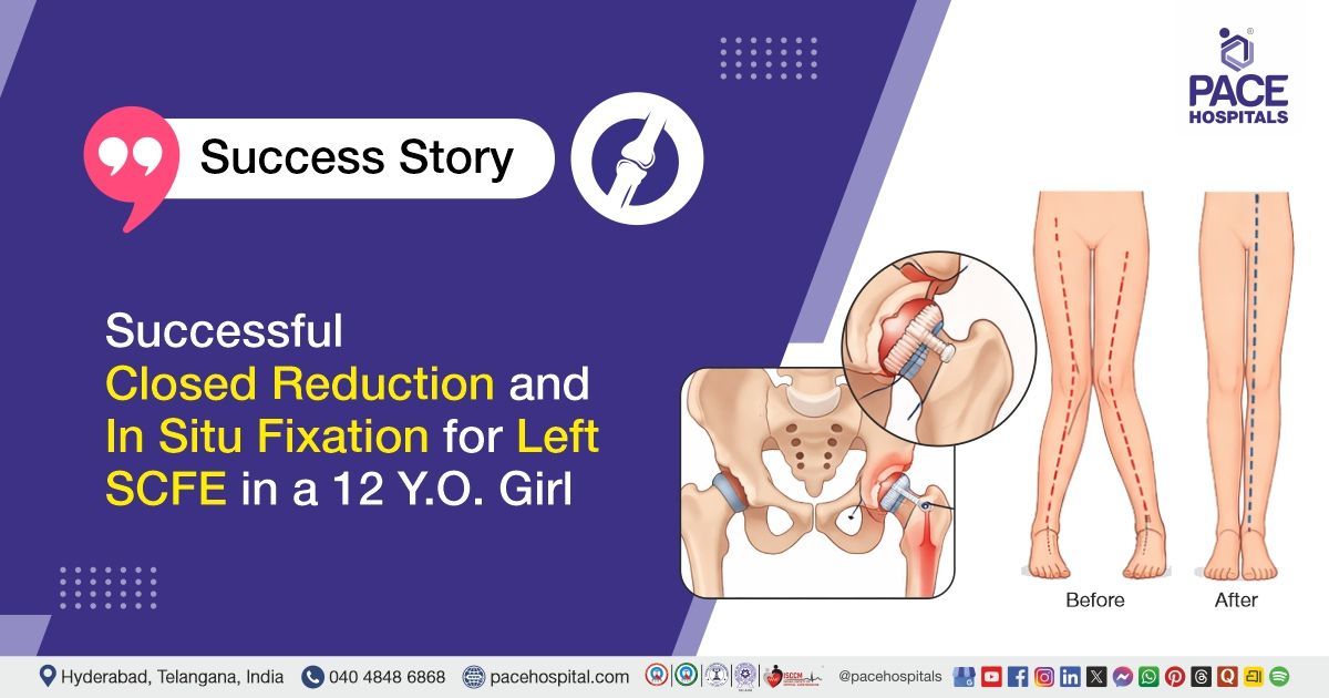
Successful Closed Reduction and In Situ Fixation for Left SCFE in a 12 Y.O. Girl
PACE Hospitals
PACE Hospitals’ expert orthopaedic team successfully performed Closed Reduction and Percutaneous In Situ Fixation using a 4 millimetre cannulated cancellous screw for left slipped capital femoral epiphysis (SCFE), followed by bilateral medial femoral hemiepiphysiodesis, in a 12-year-old female patient diagnosed with bilateral knee genu valgus deformity with left lower limb shortening and stable Grade 1 left SCFE, with the aim of stabilizing the SCFE, preventing further displacement, correcting the angular deformity of the knees, and improving limb alignment and length for better functional outcomes.
Chief Complaints
A 12-year-old female patient with a body mass index (BMI) of 18 presented to the Orthopaedic Department at PACE Hospitals, Hitech City, Hyderabad, with complaints of left hip pain, associated with difficulty in walking on the left lower limb and restricted movements in both knee joints.
Past Medical History
The patient had no significant past medical history. There was no history suggestive of chronic illnesses, prior hospitalizations, or known drug allergies. She had not been diagnosed with any systemic or metabolic disorders prior to the current presentation.
On Examination
On examination, the patient was conscious, coherent, and oriented. Bilateral genu valgus deformity was noted, more pronounced on the left side. There was tenderness over the anterior joint line of the left hip, and the patient exhibited an antalgic gait. Drehmann sign was positive. Movements of the left hip were restricted in internal rotation, flexion, and abduction. Left lower limb shortening was observed. Distal neurovascular status was normal. Vital signs were stable, and systemic examination was otherwise within normal limits.
Diagnosis
Following the clinical evaluation, the Orthopaedics team at PACE Hospitals conducted a detailed assessment focusing on the patient’s complaints of left hip pain, difficulty walking on the left lower limb, and restricted movements in both knees. The patient had a history of progressive deformity and limb shortening, which contributed to functional limitations.
A comprehensive clinical and systemic examination was performed to assess the severity of the condition. Examination revealed bilateral genu valgus deformity, more pronounced on the left side, tenderness over the anterior joint line of the left hip, antalgic gait, positive Drehmann sign, restricted internal rotation, flexion and abduction of the left hip, and left lower limb shortening. Distal neurovascular status was normal. Imaging, including X-rays and CT pelvis, confirmed bilateral knee deformity with medial femoral hemi-epiphysiodesis changes and mild Grade 1 slipped capital femoral epiphysis (SCFE) of the left hip, as per the Loder classification.
Based on these findings, the patient was advised to undergo Bilateral knee: Genu Valgus Deformity with left lower limb shortening and Stable - Grade 1 - Left SCFE Treatment in Hyderabad, India, under the care of the Orthopaedic Department.
Medical Decision Making (MDM)
After a detailed consultation with Dr. Anand Agroya, Senior Orthopaedic Consultant, a comprehensive evaluation was performed to determine the most appropriate diagnostic and therapeutic approach. Considering the patient’s history of left hip pain, difficulty walking on the left lower limb, and restricted movements in both knees, along with clinical findings of bilateral genu valgus deformity, left lower limb shortening, tenderness over the left hip, antalgic gait, positive Drehmann sign, and restricted hip movements, a focused local examination and radiological assessment were undertaken to formulate an optimal treatment strategy.
Based on the clinical findings and imaging, which confirmed stable Grade 1 left slipped capital femoral epiphysis (SCFE) and bilateral knee genu valgus deformity with left lower limb shortening, it was determined that closed reduction and percutaneous in situ fixation with a 4 mm cannulated cancellous screw for left SCFE, followed by hemiepiphysiodesis (guided growth surgery) of both knees, were identified as the most suitable intervention to relieve pain, correct deformity, restore function, and enable the patient to resume daily activities.
The patient and her family members were thoroughly counselled regarding the diagnosis, need for surgery, procedure details, risks, and recovery process. Informed consent was obtained, ensuring their understanding and involvement in the treatment plan.
Surgical Procedure
Following the diagnosis, the patient was scheduled to undergo Closed Reduction and Percutaneous In Situ Fixation with a 4 mm cannulated cancellous screw for left SCFE, followed by Hemiepiphysiodesis (guided growth surgery) of both knees Surgery in Hyderabad at PACE Hospitals, under the care and supervision of the Orthopaedic Department.
The surgical procedure involved the following steps:
- Preoperative Preparation and Anaesthesia: The patient was positioned supine on the operating table. Standard aseptic precautions were taken, and the surgical site was prepared and draped. Spinal anaesthesia was administered to provide analgesia and muscle relaxation, and prophylactic antibiotics were given to reduce the risk of infection.
- Closed Reduction of Left SCFE: Under fluoroscopic guidance, gentle manipulation of the left hip was performed to achieve closed reduction of the slipped capital femoral epiphysis (SCFE). Care was taken to avoid excessive rotation or force to prevent further slip or vascular compromise. Reduction was confirmed with intraoperative imaging, ensuring proper alignment.
- Percutaneous In Situ Fixation: A 4 mm cannulated cancellous (CC) screw was inserted percutaneously across the femoral epiphysis into the metaphysis to stabilize the SCFE. Fluoroscopy was used to confirm correct screw positioning and maintenance of femoral alignment. Intraoperative findings confirmed a Grade 1 SCFE, with less than 30-degree femoral-diaphyseal angle.
- Hemiepiphysiodesis of Bilateral Femur: Small incisions were made over the medial femoral condyles bilaterally. 8-shaped titanium plates were applied to the medial sides of both femurs, each secured with four screws, to correct and gradually reduce the genu valgus deformity. Proper fixation was verified under imaging to ensure appropriate growth modulation.
- Closure: All incisions were irrigated and closed in layers using standard surgical technique. Sterile dressings were applied, and intraoperative imaging confirmed satisfactory reduction of the SCFE and correct placement of all implants.
Postoperative Care
Postoperatively, the patient experienced intermittent episodes of fever, which were managed symptomatically. X-rays of both hips (AP view) and both knees (AP and lateral views) were taken, showing satisfactory reduction and implants in situ. On postoperative day 1, dressing was performed, and partial weight-bearing mobilization was initiated. During her hospital stay, she received treatment for infection prevention, pain relief, stomach protection, and other supportive care. She was discharged in a hemodynamically stable condition with appropriate medications and follow-up advice.
Discharge Medications
Upon discharge, the patient was prescribed medications for pain relief, infection prevention, and stomach protection. Supportive care and monitoring were also advised.
Advice on Discharge
The patient was advised to maintain strict bed rest for six weeks and to avoid bearing full weight on both lower limbs during this period.
Emergency Care
The patient was informed to contact the emergency ward at PACE Hospitals in case of any emergency or development of symptoms such as fever, hip pain, swelling, or vomiting.
Review and Follow-up Notes
The patient was advised to return for a follow-up visit with the Orthopaedic Doctor in Hyderabad at PACE Hospitals after 4 days for a wound dressing change.
Conclusion
This case highlights the successful management of a stable Grade 1 slipped capital femoral epiphysis (SCFE) of the left hip with concurrent bilateral femoral hemiepiphysiodesis (guided growth surgery) for genu valgus deformity. Closed reduction and percutaneous in situ fixation were performed with satisfactory alignment and implant placement. Postoperative recovery was uneventful with appropriate symptom management, and the patient was discharged in stable condition with instructions for restricted weight-bearing and follow-up.
Multimodal Surgical Management of Pediatric SCFE with Genu Valgus
Pediatric patients with slipped capital femoral epiphysis (SCFE) and concurrent knee deformities often benefit from a combined surgical approach that addresses both hip and lower limb alignment. An orthopaedic surgeon can perform closed reduction and percutaneous in situ fixation to stabilize the hip while minimizing the risk of growth plate injury. Hemiepiphysiodesis or guided growth techniques help gradually correct angular deformities such as genu valgus while preserving future growth potential. Accurate intraoperative imaging is crucial to ensure proper implant placement and reduce complications.
Postoperative monitoring and supportive care by the orthopaedic doctor/orthopaedic surgeon are essential for early detection of complications and safe recovery. Early controlled mobilization promotes functional recovery while protecting surgical sites. Multidisciplinary planning involving the orthopaedic surgeon and other specialists enhances overall outcomes in complex pediatric orthopedic cases.
Share on
Request an appointment
Fill in the appointment form or call us instantly to book a confirmed appointment with our super specialist at 04048486868
Appointment request - health articles
Recent Articles