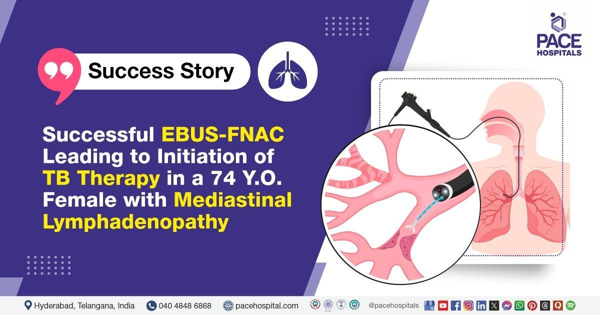
Successful EBUS-FNAC Leads to Initiation of TB Therapy in a 74-Y-O with Mediastinal Lymphadenopathy
PACE Hospitals
PACE Hospitals' expert pulmonology team successfully performed an Endobronchial Ultrasound (EBUS)-guided Fine Needle Aspiration Cytology (FNAC) on a 74-year-old female patient who had been experiencing a cold, cough with mucoid expectoration and shortness of breath (SOB) on exertion for the last 3 months. The procedure was undertaken to diagnose the condition & alleviate her symptoms, reduce the risk of future complications, and support optimal recovery.
Chief Complaints
A 74-year-old female patient with a body mass index (BMI) of 29.2 presented to the Pulmonology Department at PACE Hospitals, Hitech City, Hyderabad, with a three-month history of cold symptoms that began with a runny nose, which later progressed to nasal blockage, along with throat pain, postnasal drip, and hoarseness of voice.
She reported a persistent cough with mucoid expectoration, which was worse at night and when lying down. The patient also experienced progressive shortness of breath on exertion (mMRC grade 2–3), accompanied by wheezing and chest tightness, with symptoms worsening at night and in cold weather. There was no history of fever, loss of appetite, weight loss, or regurgitation. She reported joint pain in both wrists that improved by evening. She was fully vaccinated against COVID-19, having received two doses and a booster.
Past Medical History
She was a known case of hypertension and was on regular antihypertensive medication. She also had a history of hypothyroidism and was receiving appropriate treatment. Additionally, she had been diagnosed with asthma and was undergoing medical management.
On Examination
On the general examination, the patient was conscious and oriented, with no signs of respiratory distress. There was no peripheral lymphadenopathy or paranasal sinus tenderness. The posterior pharyngeal wall appeared within normal limits. Vital signs were stable, and the patient was afebrile.
On systemic examination, cardiovascular assessment revealed normal heart sounds. Chest examination showed bilateral crepitations along with scattered rhonchi. The abdomen was soft, with normal peristalsis.
Diagnosis
After the initial examination, the pulmonology team conducted a thorough evaluation, reviewing the patient’s medical history and symptoms. They aimed to identify any underlying causes of her respiratory issues and planned further tests to assess her symptoms.
An HRCT chest, performed outside at another hospital, revealed bilateral pneumonia along with mediastinal lymphadenopathy. The patient was admitted for further evaluation and management. Initial investigations showed normal renal function, complete blood picture, coagulation profile (PT, INR, APTT), and thyroid function. Mild hypoalbuminemia and an elevated erythrocyte sedimentation rate (ESR) were noted, while C-reactive protein (CRP), serum calcium, and angiotensin-converting enzyme (ACE) levels were within normal limits. Blood glucose levels were normal, though HbA1c suggested suboptimal glycemic control.
A 2D echocardiogram revealed normal-sized cardiac chambers, no regional wall motion abnormalities, good left and right ventricular function, grade I diastolic dysfunction, mild tricuspid regurgitation, no pulmonary arterial hypertension, trivial aortic regurgitation, and no evidence of clot, embolism, or vegetations. Multiplex flu PCR and viral screening were negative. Anti-TPO and anti-CCP antibodies were within normal limits. A chest X-ray (PA view) showed an ectatic aorta with arch calcification. Both lung fields appeared clear, hila were normal, and costophrenic angles were free. Degenerative changes were noted in the visualized spine.
Based on the confirmed findings, the patient was advised to undergo
Mediastinal Lymphadenopathy with Bilateral
Pneumonia Treatment in Hyderabad, India, under the expert care of the pulmonology department, ensuring comprehensive management.
Medical Decision Making
After being admitted to PACE Hospitals, Dr. Pradeep Kiran, Consultant Interventional Pulmonologist, thoroughly reviewed the patient’s medical history and conducted a detailed clinical examination. Laboratory investigations and imaging studies, combined with clinical suspicion, indicated the need for further evaluation.
Based on the patient's symptoms and diagnostic findings, which revealed bilateral pneumonia with mediastinal lymphadenopathy likely secondary to
tuberculosis (TB), the pulmonology team determined that performing an endobronchial ultrasound (EBUS)-guided fine needle aspiration cytology (FNAC) would be the most appropriate diagnostic procedure to further evaluate and guide management of her condition.
Surgical Procedure
Following the decision, the patient was scheduled to undergo an Endobronchial Ultrasound (EBUS)-guided Fine Needle Aspiration Cytology (FNAC) in Hyderabad at PACE Hospitals under the expert supervision of the pulmonology Department.
The patient underwent a diagnostic endobronchial ultrasound (EBUS)-guided fine needle aspiration cytology (FNAC) under general anaesthesia due to clinical and radiological findings suggestive of bilateral pneumonia with mediastinal lymphadenopathy, likely secondary to tuberculosis.
During the procedure, bronchoscopy was performed first and revealed edematous airways with mucoid secretions, indicating underlying airway inflammation. EBUS imaging identified enlarged lymph nodes at station 7, showing a heterogeneous appearance, matting, necrosis, and peripheral calcifications features commonly associated with granulomatous infections such as tuberculosis. Five needle passes were taken from station 7. Station 4R lymph nodes also showed matting and peripheral calcification, from which two passes were obtained.
A Rapid On-Site Evaluation (ROSE) conducted during the procedure was suggestive of reactive lymphadenitis, indicating a benign inflammatory process rather than malignancy. Samples were submitted for comprehensive microbiological and pathological investigations, including tests for acid-fast bacilli (AFB), bacterial and mycobacterial cultures, and histopathological examination.
The final EBUS-FNAC cytology report indicated a reactive lymphoid aspirate, which supported a diagnosis of non-malignant reactive lymphadenitis, likely of tuberculous origin. These findings helped confirm the diagnosis and guide appropriate treatment.
Postoperative Care
The procedure was uneventful, and the patient was extubated on the table before being transferred to the Medical Intensive Care Unit (MICU) for observation. After six hours of stable vital signs and satisfactory recovery, she was moved to the ward. During her hospital stay, the patient received intravenous antibiotics, oral medications, nebulization, low-flow oxygen support, antihypertensives, thyroid supplementation, and other supportive care. The EBUS cell block report is still awaited. Both the bronchoalveolar lavage (BAL) and EBUS-FNAC samples tested negative for Mycobacterium tuberculosis on acid-fast bacilli (AFB) smear and GeneXpert, though the BAL fungal smear showed a few budding yeasts.
Despite these negative microbiological results, CT and EBUS imaging revealed lymph node matting with necrosis, findings suggestive of tuberculosis. After discussing the results with the patient and her attendants, anti-tubercular therapy (ATT) was initiated according to RNTCP guidelines, based on her weight of 67.5 kg. BAL and EBUS-FNAC culture reports are still pending and will help confirm the diagnosis and guide further treatment. The patient was discharged in a relatively stable condition.
Discharge Medications
Upon discharge, the patient was prescribed a combination of medications, including fixed-dose anti-tubercular therapy, a second-line anti-tubercular agent, and an additional anti-tubercular drug for the intensive phase of treatment. She was also advised to take a vitamin B6 supplement to prevent neuropathy associated with anti-TB medications. In addition, a short course of broad-spectrum antibiotics was prescribed, along with nebulized inhalation therapy containing a bronchodilator and corticosteroid. An expectorant/mucolytic agent was included to help with respiratory secretions.
The patient was advised to continue her existing antihypertensive combination therapy and thyroid hormone replacement. Antifungal medication was also prescribed for a limited duration based on fungal smear findings.
Advice on Discharge
The patient was advised to maintain proper cough etiquette and stay well hydrated. She was informed about common side effects of anti-tubercular therapy (ATT), such as nausea, vomiting, red/orange urine, mild joint pain, and itching without rash, which can be managed with supportive medications if needed. She was also warned about serious side effects requiring medical attention, including jaundice, severe joint pain with swelling, rashes, red spots on the skin, enlarged lymph nodes, and vision problems. Regular monitoring with Liver Function Tests and Serum Uric Acid every two weeks during the first two months was also recommended.
Dietary Advice
The patient was advised to follow a low-salt, high-protein diet, which included the consumption of at least two eggs per day to support nutritional needs during treatment and recovery.
Emergency Care
The patient was informed to contact the emergency ward at PACE Hospitals in case of any emergency or development of symptoms like fever, abdominal pain, persistent cough, and vomiting.
Review and Follow-up Notes
The patient was advised to return for a follow-up visit with the Pulmonologist in Hyderabad at PACE Hospitals, after 5 days.
Conclusion
This case highlights the importance of comprehensive diagnostic workups in patients with suspected tuberculosis and mediastinal lymphadenopathy. Despite negative initial test results, imaging and clinical findings guided the decision to start anti-tubercular therapy. Early intervention, supportive care, and patient education contributed to clinical stability. Regular follow-up and monitoring were advised to ensure treatment, response and safety.
The Role of Endobronchial Ultrasound (EBUS) in Diagnosing Tuberculous Mediastinal Lymphadenopathy
Endobronchial Ultrasound (EBUS) has emerged as a crucial diagnostic modality in evaluating unexplained mediastinal lymphadenopathy. By combining bronchoscopy with real-time ultrasound guidance, EBUS enables precise sampling of deep-seated lymph nodes through fine needle aspiration cytology (FNAC). In this case, EBUS helped identify characteristic features of granulomatous inflammation, such as necrosis and calcification, highly suggestive of tuberculosis. The procedure allowed for minimally invasive, targeted tissue sampling without surgical intervention. Guided by EBUS findings, the Pulmonologist / Pulmonology doctor was able to initiate timely anti-tubercular therapy, ensuring accurate diagnosis, appropriate treatment, and better patient outcomes.
Share on
Request an appointment
Fill in the appointment form or call us instantly to book a confirmed appointment with our super specialist at 04048486868
Appointment request - health articles
Recent Articles