Successful Achilles Tendon Repair with Haglund Deformity Excision for Left Achilles Tendon Tear
PACE Hospitals
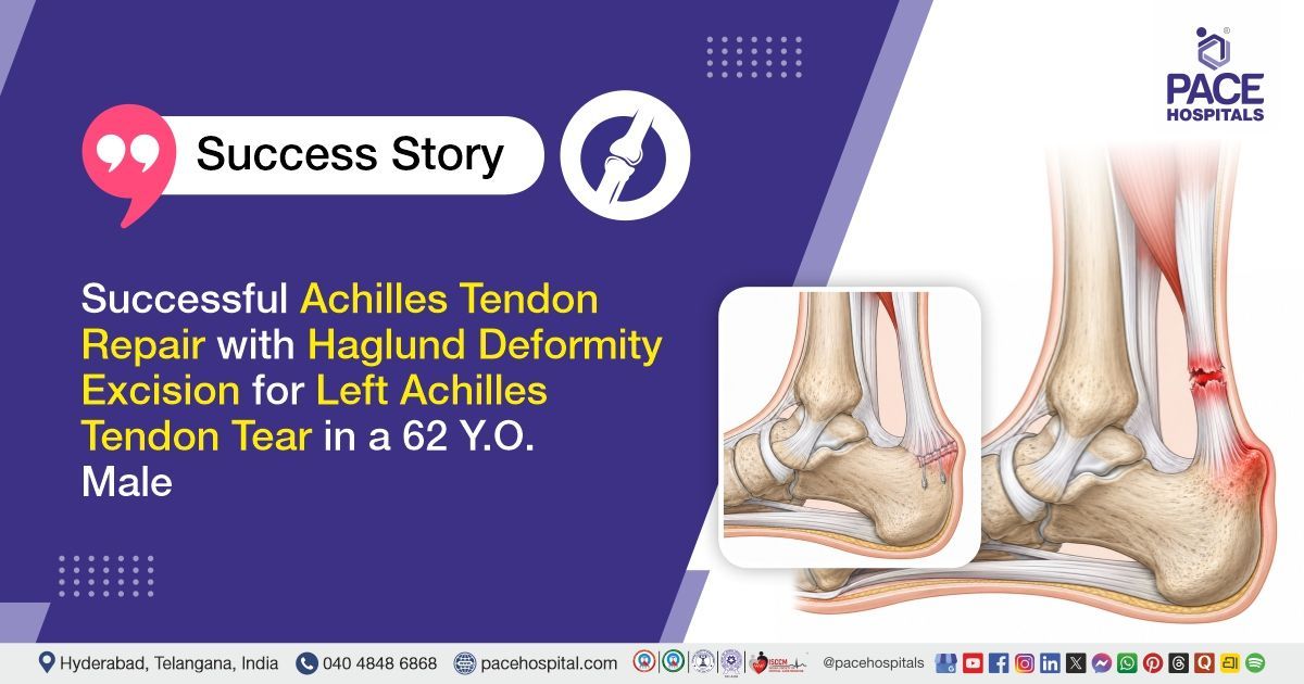
PACE Hospitals’ expert orthopaedic team successfully performed Haglund Deformity Excision along with Tendo Achilles Repair using two-anchor sutures and fiber tape in a 62-year-old male patient diagnosed with a left Tendo Achilles tear with Haglund deformity. The aim of the procedure was to remove the bony prominence causing irritation, repair the torn Achilles tendon, restore tendon strength, reduce pain, and help the patient regain better ankle movement and walking ability.
Chief Complaints
A 62-year-old male patient with a body mass index (BMI) of 22 presented to the Orthopaedic Department at PACE Hospitals, Hitech City, Hyderabad, with complaints of inability to move the left ankle for 6 days, associated with weakness of the left ankle and difficulty in walking. He also reported intermittent pain in the left ankle for the preceding two months.
Past Medical History
The patient was a known case of hypertension and was on regular antihypertensive treatment. There was no history of known drug allergies, and no other significant past medical or surgical history was documented.
On Examination
The patient was conscious, coherent, and oriented, with stable vital signs and hemodynamic status. Local examination of the left ankle revealed tenderness over the posterior aspect at the insertion of the Achilles tendon, along with a palpable gap suggestive of tendon rupture. Clinical tests assessing Achilles tendon integrity, including the Matles and Thompson tests, were positive. Increased passive dorsiflexion of the ankle was noted. Distal neurovascular examination was normal, with no evidence of vascular compromise or neurological deficit in the affected limb.
Diagnosis
Following the clinical evaluation, the Orthopaedic team at PACE Hospitals conducted a detailed assessment of the patient’s complaints of inability to move the left ankle associated with weakness, difficulty in walking, and a history of intermittent pain over the left ankle.
A thorough clinical examination revealed tenderness over the posterior aspect of the left ankle at the Achilles tendon insertion site, along with a palpable gap in the tendon region suggestive of Achilles tendon rupture. Clinical tests, including the Matles test and Thompson test, were positive, and increased passive dorsiflexion of the ankle was noted. Distal neurovascular examination was intact, and the patient was hemodynamically stable with no significant systemic abnormalities. These clinical findings were consistent with a left Achilles tendon tear associated with Haglund deformity.
Based on these findings, the patient was advised to undergo
Left Achilles Tendon Tear with Haglund Deformity Treatment in Hyderabad, India, under the care of the Orthopaedic Department to restore tendon continuity, improve ankle function and mobility, relieve pain, and prevent further functional impairment.
Medical Decision Making (MDM)
After a detailed evaluation by Dr. Anand V Agroya, Consultant Orthopaedic Surgeon at PACE Hospitals, the patient was assessed for inability to move the left ankle associated with weakness, difficulty in walking, and a history of intermittent pain over the left ankle. Clinical examination revealed tenderness over the posterior aspect of the left ankle at the Achilles tendon insertion, along with a palpable gap suggestive of tendon rupture. Clinical examination tests including the Thompson test and Matles test were positive, and increased passive dorsiflexion of the ankle was noted. Distal neurovascular status was intact, and the patient was hemodynamically stable.
Based on the clinical findings, it was determined that Left Haglund Deformity Excision with Achilles Tendon Repair using two suture anchors and FiberTape augmentation was the most appropriate management approach. This decision was made with the intent to restore tendon continuity, re-establish ankle stability and function, relieve pain, correct the underlying bony deformity, and prevent complications such as re-rupture and long-term functional impairment.
The patient and his family members were counselled regarding the diagnosis, the planned surgical procedure, possible risks and benefits, postoperative immobilization in plantar flexion, non-weight-bearing protocol, rehabilitation plan, expected recovery course, and the importance of regular follow-up for optimal functional outcome.
Surgical Procedure
Following the diagnosis, the patient was scheduled for Left Haglund Deformity Excision with Achilles Tendon Repair in Hyderabad at PACE Hospitals, using two suture anchors and FiberTape augmentation under the care and supervision of the Orthopaedic Department.
The procedure involved the following steps:
- Anaesthesia and Patient Positioning: The patient was taken to the operating room and placed in the prone position after administration of spinal anaesthesia. The left lower limb was prepared and draped under strict aseptic precautions, and a tourniquet was applied as per standard protocol.
- Surgical Exposure and Exploration: A posterior longitudinal incision was made over the left heel region centered over the Achilles tendon. Layer-wise dissection was performed to expose the tendo-Achilles. The tendon was carefully explored, revealing a near-total rupture with unhealthy and degenerated tendon margins.
- Debridement and Preparation of Tendon Ends: The torn and non-viable tendon edges were debrided to healthy tissue margins. The surrounding fibrotic and inflamed tissues were cleared to facilitate proper tendon approximation and healing.
- Haglund Deformity Excision and Tendon Repair: The posterosuperior calcaneal prominence (Haglund deformity) was excised to reduce mechanical irritation. The Achilles tendon was then repaired using suture anchors inserted into the calcaneum, and FiberTape sutures were used to achieve strong reinforcement and secure re-approximation of the tendon ends.
- Wound Closure and Immobilization: Hemostasis was achieved, and the surgical wound was closed in layers. A sterile dressing was applied, followed by a below-knee plaster of Paris slab in approximately 30 degrees of plantar flexion to protect the repair. The limb was elevated postoperatively, and the patient was shifted to recovery in stable condition.
Postoperative Care
Postoperatively, the patient had an uneventful recovery with stable vital signs and no immediate complications. The surgical site showed proper alignment and fixation of the left Achilles tendon repair, with titanium anchors in situ confirmed on radiological evaluation.
The patient received appropriate pain management, intravenous antibiotics, and supportive care. Limb elevation and anterior slab immobilization in 30° plantar flexion were maintained to protect the repair and promote healing.
Regular monitoring included assessment of distal neurovascular status, pain control, ankle mobility, and wound condition. The patient remained clinically stable throughout the hospital stay and was discharged in stable condition with instructions for non-weight-bearing mobilization, limb elevation, wound care, and follow-up consultations.
Discharge Medications
Upon discharge, the patient was prescribed medications for infection prevention, pain and inflammation control, support of recovery to reduce post-operative swelling, and gastric protection to prevent irritation associated with post-operative treatment. These medications were advised for short-term use in appropriate dosing schedules as part of routine post-operative care following Achilles tendon repair surgery.
Advice on Discharge
The patient was advised to keep the plaster dry and avoid any exposure to water. Limb elevation was recommended to help reduce swelling. Strict non-weight-bearing mobilization using a walker was advised until further instructions. The patient was also instructed to continue upper body and right lower limb exercises as previously advised.
Emergency Care
The patient was advised to contact the emergency ward at PACE Hospitals immediately in case of any emergency or if symptoms include excessive pain, swelling, redness, wound discharge, or any other signs of worsening at the surgical site.
Review and Follow-up Notes
The patient was advised to return for follow-up with the Orthopaedic Doctor in Hyderabad at PACE Hospitals after 3 days for dressing review. Suture removal was planned 15 days following the surgery.
Conclusion
This case highlights a left Achilles tendon tear associated with Haglund deformity, presenting with ankle weakness and difficulty in walking. Clinical findings were consistent with Achilles tendon rupture and confirmed on examination. The condition was managed surgically with deformity excision and tendon repair using suture anchors and FiberTape augmentation. The postoperative recovery was uneventful, and the patient was discharged in a stable condition with immobilization and follow-up advice.
Clinical Approach and Management of Achilles Tendon Rupture
Achilles tendon rupture is usually diagnosed through clinical evaluation, with bedside tests such as the Thompson and Matles tests. Imaging is usually used as an adjunct for confirmation or preoperative planning rather than for initial diagnosis. The condition may be associated with underlying degenerative changes or mechanical factors, which should be evaluated by the Orthopaedic Doctor / Orthopaedic Surgeon for comprehensive management. Treatment options include conservative functional rehabilitation or surgical repair, depending on patient factors and the severity of rupture.
Surgical techniques aim to restore tendon continuity and biomechanical strength using appropriate fixation methods. Postoperative immobilization in plantar flexion and strict non-weight-bearing are essential to protect the repair and support healing. Structured rehabilitation under the guidance of the Orthopaedic Doctor is crucial for restoring strength, mobility, and optimal functional recovery.
Frequently Asked Questions (FAQs)
Why was surgery needed for an Achilles tendon tear with Haglund deformity?
Surgery was needed because the patient had a near-total tear of the Achilles tendon along with a bony prominence at the back of the heel. This condition can affect walking, ankle movement, and tendon strength. Surgery aimed to remove the irritating bony prominence, repair the damaged tendon, reduce pain, and support better ankle function during recovery.
What is the purpose of Haglund deformity excision in this case?
Haglund deformity excision is performed to remove the extra bone at the back of the heel. This bony bump can irritate the Achilles tendon and surrounding soft tissue, leading to pain and inflammation. By taking away this prominence, pressure on the tendon is reduced, and it allows the area to heal more comfortably after Achilles tendon repair.
Why is plaster care important after this surgery?
The plaster helps keep the ankle in a safe position and protects the repaired tendon. It should be kept dry and clean to avoid skin problems, wound issues, or infection risk. The patient should avoid putting pressure on the plaster or operative site unless specifically advised by the treating orthopaedic surgeon.
When can sutures be removed after Achilles tendon repair?
Suture removal is usually planned after the wound has healed well, commonly around two weeks after surgery, depending on the doctor’s assessment. The surgeon checks the wound for healing, swelling, discharge, or infection before removing sutures. Patients should attend follow-up visits as advised and should not remove or disturb sutures on their own.
What precautions should be followed at home after discharge?
The operated leg should be kept elevated as advised to reduce swelling and discomfort. The plaster should not be made wet, and the patient should avoid bearing weight on the operated leg. Exercises for the upper body and the unaffected leg may be continued if advised, while the operated ankle should be protected until further review.
Why was Achilles tendon repair done using anchor sutures and fiber tape?
Anchor sutures and fiber tape were used to hold the torn Achilles tendon firmly to the heel bone. This gives good support to the repaired tendon while it starts healing. In the early recovery period, the tendon is still delicate, so strong fixation helps protect the repair from extra strain. It also helps keep the tendon in the right position, which is important for better ankle movement and walking function later.
Why was the foot kept in a downward position after surgery?
After Achilles tendon repair, the foot is kept slightly pointed downward so the repaired tendon is not pulled tightly. This helps the tendon heal in a safe position and protects the surgical repair in the early stage. It also reduces pressure on the operated area and supports smoother recovery. The foot position should be maintained as advised by the orthopaedic surgeon.
Why is non-weight-bearing walking advised after Achilles tendon repair?
Non-weight-bearing walking means the patient should not put body weight on the operated leg until the doctor advises. After Achilles tendon repair, the tendon needs time to heal properly without pressure. Using a walker or crutches helps the patient move around safely while protecting the repaired tendon. This also helps reduce the risk of strain, pain, or injury during the early recovery period.
How long does it usually take to recover after Achilles tendon repair with Haglund deformity excision?
Recovery time can vary from person to person. It depends on the patient’s age, tendon condition, wound healing, and rehabilitation progress. In the first few weeks, the main focus is on protecting the repaired tendon and allowing the surgical wound to heal properly. Walking, ankle movement, and strengthening exercises are started gradually only after the surgeon’s review.
When should urgent medical care be taken after Achilles tendon surgery?
Urgent medical care is needed if the patient develops fever, increasing pain, swelling, wound discharge, bad smell from the wound, tight plaster, numbness, toe colour change, or sudden worsening of symptoms. These signs may point to wound infection, pressure, or blood circulation problems. Early medical review helps identify the problem quickly and prevents further complications.
Share on
Request an appointment
Fill in the appointment form or call us instantly to book a confirmed appointment with our super specialist at 04048486868
Appointment request - health articles
Recent Articles