Successful Bifrontal Craniotomy & Anterior Cranial Fossa Base Repair for Right Ethmoidal Encephalocele
PACE Hospitals
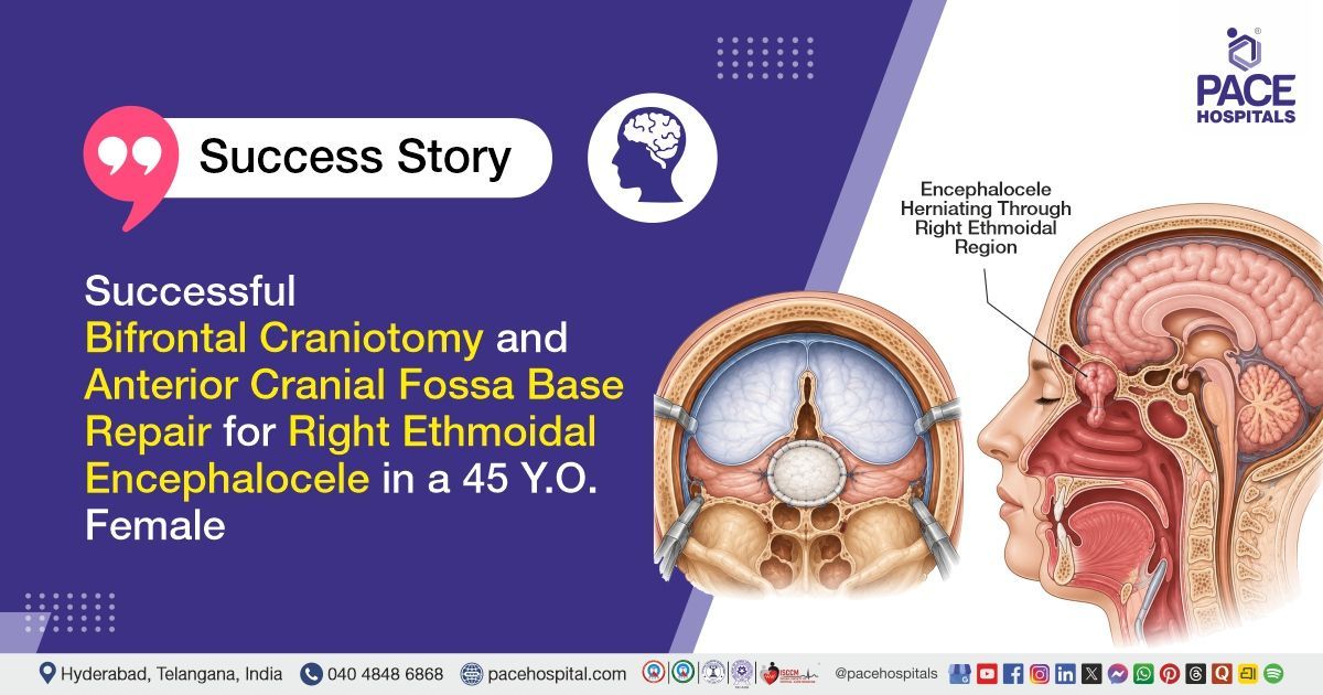
PACE Hospitals’ expert Neurosurgery team successfully performed a Bifrontal Craniotomy, Excision of Encephalocele, and Anterior Cranial Fossa Base Repair on a 45-year-old female patient diagnosed with right ethmoidal encephalocele with chronic CSF rhinorrhoea. The primary aim of the procedure was to remove the herniated brain tissue, repair the skull base defect, stop the cerebrospinal fluid (CSF) leak, prevent recurrent infections such as meningitis, and restore the integrity of the anterior cranial fossa.
Chief Complaints
A 45-year-old female patient with a body mass index (BMI) of 21 presented to the Neurosurgery Department at PACE Hospitals, Hitech City, Hyderabad, with complaints of intermittent watery nasal discharge for the past two and a half years. She reported experiencing similar symptoms four years earlier, with recurrent episodes of clear nasal discharge occurring on and off.
Past Medical History
The patient was a known case of Type 2 Diabetes Mellitus and was on regular antidiabetic medication. She also had a history of seizure disorder and was receiving antiepileptic treatment. There was a history of recurrent episodes of clear watery nasal discharge over the preceding several years, accompanied by intermittent fever and loss of smell (anosmia), suggestive of a chronic cerebrospinal fluid (CSF) leak. No prior history of limb weakness, visual disturbances, speech impairment, or other significant neurological deficits was reported.
On Examination
On examination, the patient was moderately built and nourished with stable vital signs. Cardiovascular and respiratory system examinations were within normal limits. Neurological examination revealed anosmia (loss of smell sensation), but there were no signs of meningitis. Motor power was normal in all four limbs, with no focal neurological deficits. There was no evidence of limb weakness, visual disturbances, or speech abnormalities. Overall, the systemic examination was normal except for the loss of smell sensation.
Diagnosis
Upon admission to PACE Hospitals, the patient underwent a comprehensive clinical evaluation along with a detailed review of her medical history and preoperative investigations conducted by the Neurosurgery team.
Laboratory investigations revealed mild microcytic hypochromic anemia with evidence of iron deficiency. Renal function tests, liver function tests, serum electrolytes, thyroid profile, coagulation parameters, and viral screening were largely within normal limits. Diabetes evaluation showed poorly controlled blood glucose levels with elevated HbA1c. Inflammatory markers were elevated, while cardiac assessment demonstrated preserved ventricular function with mild diastolic dysfunction.
Neuroimaging demonstrated a right ethmoidal encephalocele associated with a defect in the cribriform plate and chronic cerebrospinal fluid (CSF) rhinorrhoea. Clinically, the patient had a long-standing history of intermittent watery nasal discharge, recurrent febrile episodes, seizures on medication, and loss of smell sensation, with no focal motor deficits or signs of meningitis.
Based on the confirmed diagnosis, the patient was advised to undergo Right Ethmoidal Encephalocele with Chronic CSF Rhinorrhoea Treatment in Hyderabad, India under the expert care of the Neurosurgery Department.
Medical Decision-Making (MDM)
After a detailed consultation with Dr. U. L. Sandeep Varma (Consultant Neurosurgeon), and cross-consultation with Dr. Tripti Sharma (Consultant Endocrinologist), a comprehensive evaluation was conducted focusing on the patient’s presentation of chronic watery nasal discharge for more than two and a half years, recurrent fever episodes, seizures on medication, and loss of smell sensation suggestive of a cerebrospinal fluid leak.
Clinical examination, laboratory investigations including complete blood picture, renal and liver function tests, serum electrolytes, coagulation profile, viral markers, chest X-ray, CT brain imaging, and preoperative systemic assessment were reviewed. Findings confirmed a right ethmoidal encephalocele with chronic cerebrospinal fluid (CSF) rhinorrhoea secondary to a defect in the right cribriform plate, associated with herniation of intracranial contents through the anterior skull base. The patient was also noted to have diabetes mellitus under medical management, while no major cardiopulmonary contraindications were identified for surgery.
It was determined that bifrontal craniotomy with excision of the encephalocele and anterior cranial fossa base repair under general anesthesia was the most appropriate intervention to excise the herniated gliotic brain tissue, repair the skull base defect, arrest the chronic CSF leak, reduce the risk of recurrent meningitis and intracranial infections, prevent further neurological complications, and achieve durable reconstruction of the anterior cranial fossa.
The patient and family were counselled in detail regarding the diagnosis, surgical procedure, perioperative risks and benefits, postoperative care including CSF diversion through lumbar drainage, wound care, activity restrictions, seizure management, blood sugar control, medication adherence, warning signs of recurrence or infection, and the importance of regular follow-up to optimize recovery and long-term outcomes.
Surgical Procedure
Following the decision, the patient was scheduled to undergo Bifrontal Craniotomy with Excision of the Encephalocele and Anterior Cranial Fossa Base Repair Surgery in Hyderabad at PACE Hospitals under the expert supervision of the Neurosurgery Department.
The procedure involved the following steps:
- Anesthesia and Positioning with Lumbar Drain Placement: The patient was administered general anesthesia (GA) and positioned appropriately for bifrontal cranial access. A lumbar drain was inserted under sterile precautions to facilitate cerebrospinal fluid (CSF) diversion and reduce intracranial pressure during the procedure.
- Surgical Exposure via Bicoronal Incision and Craniotomy: A bicoronal scalp incision was made under strict aseptic precautions. A pedicled pericranial flap was carefully harvested for later reconstruction. A bifrontal craniotomy was performed after exposing the cranial vault, and the dura was incised at the skull base following ligation of the superior sagittal sinus to access the anterior cranial fossa.
- Identification and Excision of Encephalocele: The right-sided ethmoidal encephalocele was identified at the skull base defect. The herniated gliotic brain tissue was carefully cauterized and excised to prevent recurrence and eliminate the source of CSF leakage.
- Skull Base Defect Repair and Reconstruction: The defect in the right cribriform plate was identified and its margins were fully exposed. The defect was repaired using layered reconstruction, including temporalis muscle and fascia graft placement, application of dural substitute and reinforcement with a pedicled pericranial flap to achieve a watertight closure of the anterior cranial fossa floor.
- Closure and Stabilization: The frontal sinus was packed and exteriorized using the pericranial flap technique. The bone flap was repositioned and securely fixed. The dura was closed, and the surgical wound was closed in layers over a surgical drain to prevent fluid collection and ensure proper healing.
Postoperative Care
Postoperatively, the patient was managed in the intensive care unit (ICU) with cerebrospinal fluid diversion through a lumbar drain at controlled rates to support skull base healing and prevent recurrence of CSF leak, along with close neurological monitoring for seizures, infection, and rhinorrhoea. She was shifted to the ward after stabilization, and the lumbar drain was removed on postoperative day 2 following adequate recovery.
Early ambulation was initiated on postoperative day 3 with regular wound care to ensure proper healing and prevent complications. Blood glucose levels were monitored and optimized due to underlying diabetes mellitus, and seizure prevention was continued given her clinical history.
Postoperative CT brain showed bilateral frontal craniotomy and anterior cranial fossa repair changes, with a residual CSF-density sac suggestive of persistent meningocele/residual CSF leak and focal right basal frontal hemorrhagic contusions. The postoperative course remained uneventful, and the patient was discharged in stable condition with advice on activity restriction and regular follow-up.
Discharge Medications
Upon discharge, the patient was prescribed medications for prevention of postoperative infection following skull base surgery, anaerobic infection prevention related to sinus and intracranial involvement, and seizure prevention in view of the pre-existing seizure disorder and recent neurosurgical intervention.
Additional medications were given to reduce cerebrospinal fluid production and support healing of the skull base repair, provide gastric protection during recovery, control postoperative pain, support neurological recovery, regulate bowel movements to avoid straining, maintain optimal blood sugar control to promote wound healing and reduce infection risk, provide symptomatic relief for cough, and ensure local wound site protection to prevent surgical site infection.
Advice on Discharge
The patient was advised to avoid coughing, sneezing, straining, and excessive physical activity. She was instructed to follow proper wound care on alternate days. Regular blood sugar monitoring was also advised.
Emergency Care
The patient was instructed to contact the emergency ward at PACE Hospitals if she developed fever, seizures, recurrent nasal discharge suggestive of cerebrospinal fluid leak, severe headache, or weakness of limbs.
Review and Follow-up Notes
The patient was advised to return for follow-up with the Neurosurgeon in Hyderabad at PACE Hospitals after 10 days for postoperative review and wound inspection.
Conclusion
This case highlights a right ethmoidal encephalocele with chronic cerebrospinal fluid rhinorrhoea managed by bifrontal craniotomy, excision of encephalocele, and anterior cranial fossa base repair. Postoperatively, the patient showed good recovery with complete cessation of nasal discharge and stable neurological status. No signs of meningitis or new neurological deficits were noted. Overall, the patient had a favorable surgical outcome with uneventful recovery.
Complex Skull Base Reconstruction in Anterior Cranial Fossa Defects
Anterior cranial fossa skull base defects with cerebrospinal fluid leakage are complex conditions that require a structured surgical approach by a Neurosurgeon/Neurosurgery doctor. Management generally involves secure closure of the defect using multilayer reconstruction techniques to restore the barrier between the intracranial and sinonasal spaces. Reduction of cerebrospinal fluid pressure through temporary diversion methods may be used to support healing of the repair site. The primary goal is to prevent ongoing leakage and reduce the risk of intracranial infections. Long-term monitoring is important to ensure the integrity of the repair and to detect any recurrence early. With timely intervention, most cases have good functional outcomes and reduced risk of serious complications
Frequently Asked Questions (FAQs)
Why was surgery needed for chronic CSF leakage from the nose?
Surgery was required because the patient had watery nasal discharge for nearly two and a half years, which was due to leakage of cerebrospinal fluid from the skull base. Long-standing CSF leakage can increase the risk of repeated fever, meningitis, seizures, and other serious neurological complications.
What does right ethmoidal encephalocoele with CSF rhinorrhoea mean?
Right ethmoidal encephalocoele with CSF rhinorrhoea refers to an abnormal defect in the skull base in the region of the right ethmoid sinus, through which intracranial contents such as meninges or brain tissue may herniate. This defect also creates an abnormal communication between the intracranial space and the nasal cavity, leading to leakage of cerebrospinal fluid (CSF) through the nose, which presents as clear, watery nasal discharge. It is not a normal nasal condition and usually indicates an underlying skull base defect. The condition typically requires evaluation and surgical management by a neurosurgeon/neurosurgery doctor to prevent complications such as recurrent infections and other intracranial risks.
Why was a bifrontal craniotomy performed in this case?
Bifrontal craniotomy was done to give the neurosurgeon safe and clear access to the anterior skull base. This helped in identifying the skull base defect, removing the encephalocoele, and repairing the leakage site properly.
How does anterior cranial fossa base repair stop CSF leakage?
The repair closes the opening in the skull base from where the CSF was leaking. In this case, tissue layers were used to seal and strengthen the defect, helping prevent further leakage of fluid into the nose.
Why was a lumbar drain placed after the surgery?
A lumbar drain was used to temporarily divert cerebrospinal fluid and reduce pressure on the repaired area. This gives the skull base repair time to heal and lowers the chance of the leak reopening soon after surgery.
Was the surgery successful in stopping the nasal discharge?
Yes. At the time of discharge, the patient’s post-nasal drip and nasal discharge had stopped. The patient was conscious, stable, and discharged with medicines, precautions, and follow-up advice.
What precautions are important after CSF leak repair surgery?
The patient should avoid coughing, sneezing forcefully, straining, heavy physical activity, and anything that increases pressure inside the head. These precautions are important because excess pressure can disturb the repaired skull base area.
Can CSF leakage come back after surgery?
CSF leakage may recur in some cases after surgery, particularly if there is delayed healing, increased intracranial pressure, or strain at the repair site. It is important to monitor for any recurrence of clear, watery nasal discharge. If such symptoms appear, medical evaluation should be sought promptly.
Why is follow-up important after encephalocoele excision and skull base repair?
Follow-up after encephalocoele excision and skull base repair is important to assess proper wound healing and overall neurological recovery. It also helps the neurosurgeon to monitor seizure control and detect any early signs of recurrent CSF leakage. Regular reviews allow timely adjustment of medications if needed. Follow-up also ensures that any complications are identified and managed at an early stage.
When should the patient seek urgent medical care after discharge?
Urgent medical care should be sought if the patient develops fever, seizures, recurrence of clear watery nasal discharge, severe headache, weakness of limbs, confusion, or any problems related to the surgical wound. These symptoms may indicate infection, recurrence of CSF leak, or other neurological complications. Early evaluation by a neurosurgeon/neurosurgery doctor is important in such situations.
Share on
Request an appointment
Fill in the appointment form or call us instantly to book a confirmed appointment with our super specialist at 04048486868
Appointment request - health articles
Recent Articles