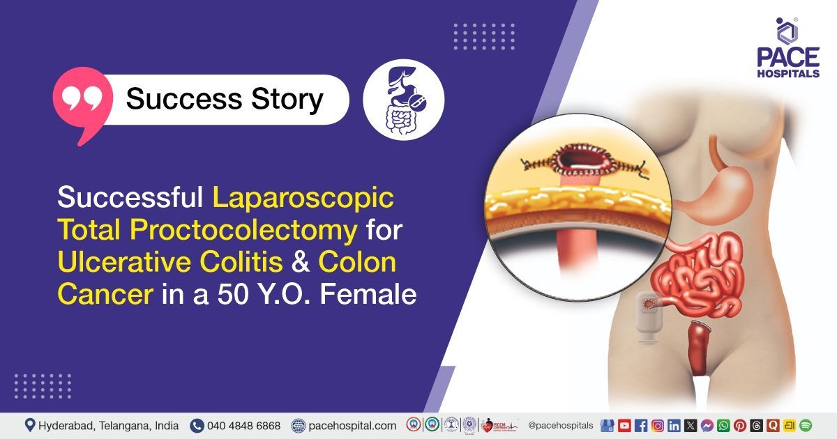
Successful Laparoscopic Total Proctocolectomy for Ulcerative Colitis & Colon Cancer in a 50 Y.O. Female
PACE Hospitals
The PACE Hospitals expert surgical gastroenterology team successfully performed a Laparoscopic Total Proctocolectomy with Ileal Pouch Anal Anastomosis (IPAA) and Covering Ileostomy (Stage-1) in a 50-year-old female patient diagnosed with ulcerative colitis (pan-colitis). Pre-operative high-grade dysplasia on recto-sigmoid biopsy progressed to moderately differentiated adenocarcinoma (pT3 N1b, 2/17 lymph nodes positive, lymphovascular invasion present) on final histopathology. The procedure aimed to remove the diseased colon and rectum, restore bowel continuity through ileal pouch creation, and improve the patient’s quality of life.
Chief Complaints
A 50-year-old female patient with a body mass index (BMI) of 18 presented to the Surgical Gastroenterology Department at PACE Hospitals, Hitech City, Hyderabad, with complaints of increased frequency of stools and passage of blood and mucus in stools for the past 5 years. She initially experienced up to 10 episodes of loose stools per day, associated with intermittent abdominal pain, urgency, and tenesmus. Although she was diagnosed with ulcerative colitis 5 years ago and managed with medications, she had multiple relapses over time. At present, she continues to have 5 to 6 bowel movements per day, often mixed with mucus, which significantly affects her quality of life.
Past Medical History
The patient was a known case of ulcerative colitis for the past 5 years and had been on medications. She had experienced approximately 10 episodes of relapse, all managed conservatively. On colonoscopic biopsy from a recto-sigmoid polyp, she was diagnosed with high-grade dysplasia. She also had a history of cervical spondylitis for 4 years and had undergone drainage of a right leg abscess 2 months prior. There were no major comorbid conditions reported. She had a past surgical history of two lower-segment cesarean sections (LSCS).
On Examination
On general examination, the patient was conscious, coherent, and hemodynamically stable. Vital parameters were within normal limits. Systemic examination did not reveal any significant abnormalities, and abdominal examination did not show acute tenderness or distension.
Diagnosis
Upon admission, the patient was thoroughly evaluated by the Surgical Gastroenterology team. She presented with a long-standing history of increased frequency of stools associated with passage of blood and mucus for the past 5 years. The symptoms were accompanied by intermittent abdominal pain, urgency, and tenesmus, with multiple episodes of relapse despite ongoing medical therapy. Recently, there was persistence of symptoms with 5–6 episodes of stools per day. Clinical history was suggestive of chronic Ulcerative Colitis with active disease.
The patient underwent further diagnostic evaluation. Colonoscopic examination with biopsy from a recto-sigmoid polyp revealed high-grade dysplasia, indicating a premalignant transformation. Laboratory investigations showed anemia with microcytic hypochromic picture and intermittent neutrophilic leukocytosis, while renal and liver function tests were within normal limits. Overall clinical, endoscopic, and histopathological findings confirmed extensive colonic involvement.
Based on these findings and the confirmed diagnosis of Ulcerative colitis (pan-colitis) with high-grade dysplasia on pre-op biopsy, confirmed as pT3 N1b adenocarcinoma on histopathology, the patient was advised to undergo advanced colon cancer treatment in Hyderabad, India, under the expert care of the Surgical Gastroenterology Department to achieve complete tumor removal, prevent disease progression, and improve long-term survival outcomes.
Medical Decision Making (MDM)
After a detailed consultation with Dr. CH Madhusudhan, Senior Consultant Surgical Gastroenterologist along with the team including Dr. Suresh Kumar S, Consultant Surgical Gastroenterologist, a comprehensive evaluation was carried out to determine the most appropriate diagnostic and therapeutic approach for the patient.
Based on the clinical and diagnostic evaluation, it was determined that Laparoscopic total proctocolectomy with ileal pouch anal anastomosis (IPAA) and covering ileostomy (Stage-1 procedure) was identified as the most appropriate treatment to remove the diseased colon and rectum, and restore bowel continuity while preserving continence.
The patient and her family members were counselled in detail regarding the diagnosis, the need for staged surgical management, the procedure involved, potential risks and complications, expected postoperative recovery, stoma care, and the plan for ileostomy closure in the subsequent stage. The benefits of surgery in improving long-term outcomes and quality of life were clearly explained before proceeding with the treatment.
Surgical Procedure
Following the decision, the patient was scheduled for Laparoscopic Total Proctocolectomy with Ileal Pouch Anal Anastomosis (IPAA) and Covering Ileostomy (Stage-1 procedure) in Hyderabad at PACE Hospitals under the expert care of the Surgical Gastroenterology Department.
The procedure involved the following steps:
- Anesthesia and Positioning: The patient was placed under general anesthesia with endotracheal intubation. She was positioned supine with appropriate padding, and a modified lithotomy position with slight Trendelenburg tilt was used to facilitate optimal access to the pelvis and abdominal cavity.
- Port Placement and Laparoscopic Access: A standard multi-port laparoscopic technique was used. Pneumoperitoneum was created using carbon dioxide to establish adequate working space. A laparoscope and working instruments were introduced through the ports to visualize the abdominal cavity, colon, and pelvic structures.
- Initial Exploration: Diagnostic laparoscopy was performed to assess the extent of disease. There was no evidence of metastatic deposits over the peritoneum, liver, or omentum, and no ascites or significant lymphadenopathy was noted. The abdominal viscera appeared normal.
- Mobilization of Colon and Rectum: The procedure began with mobilization of the sigmoid colon by incising the peritoneum at the sacral promontory. The mesocolon was carefully dissected, and the plane between the mesocolon and retroperitoneum was developed. Critical structures such as the ureter and gonadal vessels were identified and preserved.The inferior mesenteric artery and vein were dissected, ligated, and divided while preserving surrounding nerve plexuses. The dissection was continued proximally to mobilize the descending colon, splenic flexure, transverse colon, hepatic flexure, and ascending colon. The ileocolic pedicle was divided, and the ileum was transected at the ileocecal junction.
- Rectal Dissection: The rectum was mobilized circumferentially in the avascular plane, starting posteriorly, followed by lateral and anterior dissection while safeguarding adjacent structures, including the vagina. Dissection was continued down to the pelvic floor, and the rectum was divided.
- Specimen Extraction: The entire colon and rectum were removed through a small infra-umbilical incision, ensuring minimal tissue handling and preservation of surrounding structures.
- Ileal Pouch Construction (J-Pouch): A 15 cm ileal pouch was created by aligning two limbs of the ileum side-by-side and using linear staplers to form a J-shaped reservoir. This pouch serves as a substitute for the removed rectum.
- Ileal Pouch Anal Anastomosis (IPAA): A circular stapler was introduced trans-anally, and the ileal pouch was connected to the anal canal. The anastomosis was checked for integrity using an air leak test to ensure there was no leakage.
- Covering Ileostomy: A temporary diverting ileostomy was created proximal to the pouch to protect the anastomosis and allow proper healing by diverting fecal flow.
- Drain Placement and Closure: A pelvic drain was placed near the anastomosis site. Pneumoperitoneum was released, ports were removed, and all incisions were closed in layers using absorbable sutures.
Postoperative Care
The procedure was uneventful, and the patient was closely monitored during the postoperative period. Initially, she experienced nausea with one episode of vomiting on the second postoperative day, which was managed with medications to control nausea and improve gastrointestinal motility. She was maintained on fluids to prevent dehydration, medications to prevent and treat infection, pain relief, and supportive care to maintain gut health. The patient showed gradual clinical improvement, the stoma started functioning appropriately, and she was subsequently initiated on a liquid diet.
Discharge Medications
The patient was discharged in hemodynamically stable condition with a central venous catheter (right internal jugular vein) and a drain in situ. Upon discharge, the patient was prescribed medications to prevent and treat infection, reduce stomach acid, support nutrition and blood health, maintain gut health, and provide overall supportive care to promote healing and recovery.
Advice on Discharge
The patient was advised regarding proper stoma care, central line care, and wound care. She was instructed to follow a soft, high-protein diet along with adequate hydration to support recovery and maintain nutritional balance.
Emergency Care
The patient was informed to contact the emergency ward at PACE Hospitals in case of any emergency or development of symptoms such as fever, abdominal pain, vomiting, or chest pain.
Review and Follow-up Notes
The patient was advised to follow up with the Surgical gastroenterologist in Hyderabad at PACE Hospitals after 3 days. She was also counselled regarding the need for ileostomy closure (Stage-2 procedure), which is planned after 6 to 8 weeks, depending on recovery and clinical status.
Conclusion
This case highlights the successful management of a patient with long-standing ulcerative colitis complicated by malignant transformation into moderately differentiated adenocarcinoma. Following comprehensive evaluation and multidisciplinary decision-making, laparoscopic total proctocolectomy with ileal pouch anal anastomosis was performed successfully. Histopathological examination confirmed Stage III disease (pT3 N1b) with regional lymph node involvement. The patient tolerated the procedure well, showed good postoperative recovery, and was discharged in stable condition with appropriate follow-up advice. Further oncological evaluation for adjuvant therapy was recommended. This approach provided effective surgical management and significantly improved the patient’s quality of life.
Multidisciplinary Approach in Managing Ulcerative Colitis with High-Grade Dysplasia
Management of Ulcerative Colitis with high-grade dysplasia requires a comprehensive multidisciplinary approach involving a surgical gastroenterologist/surgical gastroenterology doctor, a pathologist, an anesthesiologist, and a nutrition specialist. In long-standing or complicated cases, coordinated care is essential due to the increased risk of progression to colorectal cancer. A detailed evaluation using colonoscopy with biopsy, histopathology, and laboratory investigations helps confirm the diagnosis. The identification of high-grade dysplasia is a crucial turning point, as it indicates a precancerous condition requiring definitive surgical management rather than continued medical therapy.
Surgical intervention, particularly laparoscopic total proctocolectomy with ileal pouch anal anastomosis (IPAA), is considered the gold standard in such cases, as it removes the diseased colon and eliminates cancer risk while preserving continence. Minimally invasive techniques offer benefits such as reduced pain, fewer complications, and faster recovery. Postoperative care focuses on monitoring, infection prevention, stoma care, and nutritional support. A coordinated multidisciplinary approach ensures individualized treatment, optimal outcomes, and improved quality of life.
Share on
Request an appointment
Fill in the appointment form or call us instantly to book a confirmed appointment with our super specialist at 04048486868
Appointment request - health articles
Recent Articles