Successful Laminectomy with Tumor Removal for Intradural Schwannoma
PACE Hospitals
PACE Hospitals’ expert Neurosurgery team successfully performed an L2–3 Laminectomy with Excision of a Tumor on a 50-year-old female patient diagnosed with an L2–3 intradural space-occupying lesion (schwannoma). The aim of the procedure was to relieve pressure on the spinal cord and nerve roots, alleviate symptoms, and achieve complete removal of the tumor to prevent recurrence and restore neurological function.
Chief Complaints
A 50-year-old female patient with a body mass index (BMI) of 21 presented to the Neurosurgery Department at PACE Hospitals, Hitech City, Hyderabad, with complaints of low back ache associated with bilateral lower limb paresthesias and radiculopathy for the past 6 months. She also reported urinary urge incontinence for the past 4–5 months. The symptoms were persistent and progressively bothersome. There was no history of weakness in the limbs, trauma, fever, neck stiffness, or bowel incontinence.
Past Medical History
The patient had a medical history of Diabetes Mellitus (DM) and Hypertension (HTN), both of which were managed with medication. She also had a history of a post-laparoscopic salpingectomy performed for hydrosalpinx about two months ago.
On Examination
On examination, the patient was conscious, coherent, and oriented to time, place, and person. She was moderately built and nourished. Her vitals were stable, with a normal heart rate, blood pressure, and respiratory rate. On systemic examination, there was no tenderness in the abdominal region, and the cardiovascular and respiratory systems were normal. The neurological examination showed a power of 4+/5 in both lower limbs, with normal muscle tone and flexor plantar responses.
Diagnosis
Upon admission to PACE hospitals, the patient underwent a comprehensive clinical evaluation, along with the patient’s medical history and diagnostic investigations conducted by the Neurosurgery team.
Laboratory and special investigations were conducted as part of the preoperative assessment. Complete blood counts (CBC) revealed thrombocytosis and neutrophilic leukocytosis, but no systemic infection was evident. Viral screening for HIV, Hepatitis B, and Hepatitis C was negative. Renal function tests and serum electrolytes were stable, with mild hyponatremia noted and corrected during the hospital stay. Chest X-ray demonstrated normal heart size and clear lung fields. Blood and urine cultures initially revealed bacterial growth, for which appropriate antibiotics were administered, with subsequent cultures showing no further growth.
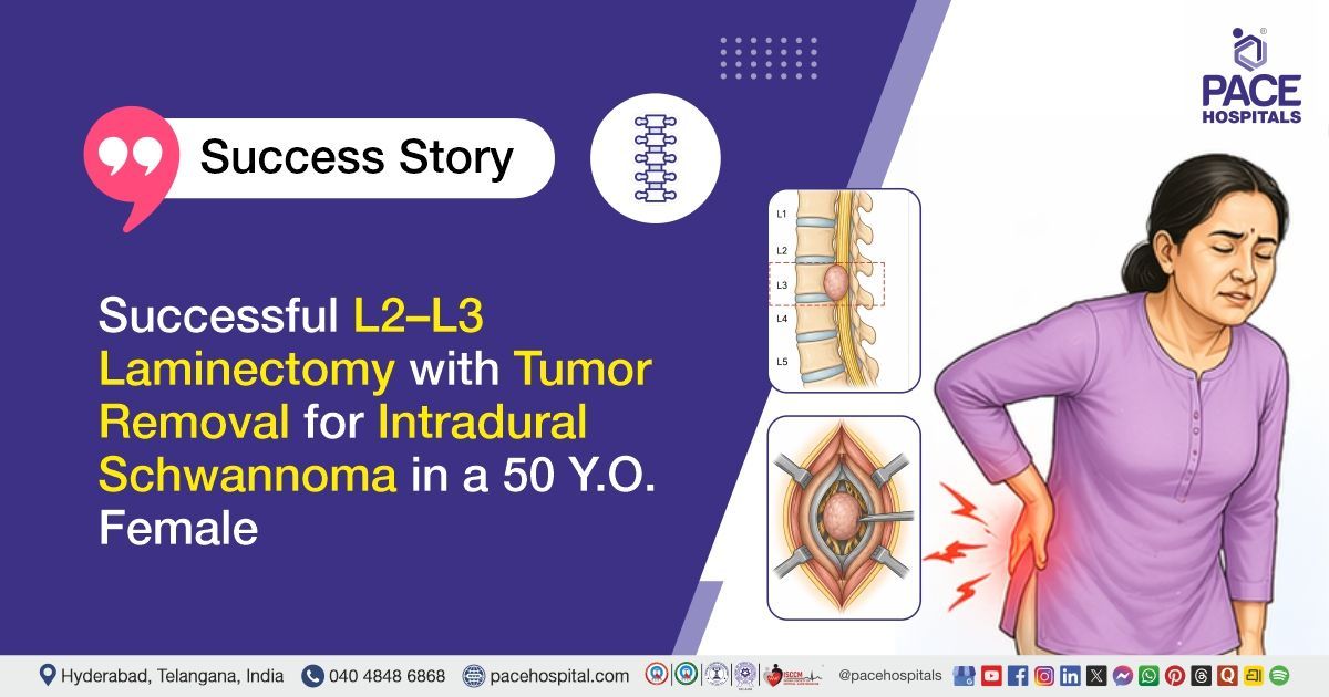
Neuroimaging investigations, including Computed Tomography (CT) and Magnetic Resonance Imaging (MRI), demonstrated a well-defined intradural extramedullary lesion at the L2-L3 levels. The lesion showed mass effect on the adjacent cauda equina nerve roots. The possibility of Schwannoma was suggested.
Based on the confirmed diagnosis, the patient was advised to undergo L2-3 Intra Dural Sol – Schwannoma Treatment in Hyderabad, India, under the expert care of the Neurosurgery Department.
Medical Decision-Making (MDM)
After a detailed consultation with Dr. U. L. Sandeep Varma, Consultant Neurosurgeon, along with cross-consultation by Dr. Govind Verma, Interventional Gastroenterologist, a comprehensive neurological assessment and radiological evaluation were performed to determine the most appropriate management and therapeutic approach. The patient presented with low back ache, bilateral lower limb paresthesias, radiculopathy, and urinary urge incontinence, and neuroimaging confirmed an L2–L3 intradural extramedullary lesion causing compression of the cauda equina nerve roots, suggestive of schwannoma.
The clinical evaluation revealed features of progressive neural compression with early bladder involvement. It was determined that L2–L3 laminectomy with excision of the intradural tumor under general anaesthesia was identified as the most appropriate surgical intervention. This procedure was aimed at complete tumor removal, decompression of neural elements, relief of symptoms, and prevention of further neurological deterioration.
The patient and her family members were counseled in detail regarding the diagnosis, surgical intervention, postoperative care, medication adherence, physiotherapy, and the need for regular follow-up to optimize long-term recovery.
Surgical Procedure
Following the decision, the patient was scheduled to undergo an L2–L3 Laminectomy Surgery in Hyderabad at PACE Hospitals along with complete excision of the intradural tumor under the expert supervision of the Neurosurgery Department.
The procedure involved the following steps:
- Patient Positioning and Preparation: Under strict aseptic precautions, the patient was administered general anaesthesia and positioned prone. The operative field was thoroughly painted and draped in a sterile manner.
- Surgical Exposure: A vertical midline incision was made over the lumbar region. The incision was deepened in layers, and the paraspinal muscles were carefully separated to expose the posterior elements of the spine.
- Laminectomy and Dural Access: L2–L3 laminectomy was performed to decompress and access the spinal canal. The dura was then carefully incised to expose the intradural space.
- Tumor Identification and Excision: A space-occupying lesion (SOL) was identified among the nerve roots, with one root/filum observed entering the lesion. The tumor, which was soft and cystic in consistency, was meticulously dissected from adjacent nerve roots and excised in a piecemeal fashion, ensuring maximal safe removal.
- Reconstruction and Wound Closure: Duroplasty was performed using 4-0 nylon sutures and reinforced with Duragen. The removed bone fragments were repositioned in the laminectomy site. A drain was placed, and the wound was closed in layers under aseptic conditions, followed by sterile dressing application.
Postoperative Care
Postoperatively, the patient was shifted to the Intensive Care Unit (ICU) for overnight observation. The surgical drain was removed on postoperative day 2, following which the patient was ambulated. She developed abdominal pain for which a medical consultation was obtained and appropriate advice was followed. The Foley’s catheter was removed after intermittent clamping. The patient was mobilized under physiotherapy guidance and received regular physiotherapy sessions. An orthopedic opinion was taken, and conservative management for bilateral knee pain was advised. The patient was planned for discharge with continuation of medications and physiotherapy as advised.
Discharge Medications
Upon discharge, the patient was prescribed medications for the prevention of postoperative infection, management of neuropathic pain and radicular symptoms, gastric protection, nerve healing and neuroregeneration, short-term anti-inflammatory and analgesic effect, and relief of dizziness and vertigo. She was also advised to undergo treatment to regulate bowel movements and prevent constipation, along with supportive therapy for gastrointestinal comfort. In addition, medications were continued for long-term control of hypertension and diabetes mellitus as per her existing medical condition, with instructions for regular adherence and follow-up.
Advice on Discharge
The patient was advised that she may take a bath and resume routine activities such as walking and climbing stairs as tolerated. She was instructed to avoid strenuous exercises and heavy lifting. Continuation of physiotherapy as advised was recommended to support recovery and improve functional outcomes. The patient was also advised to strictly follow the prescribed medications and attend regular follow-up visits as scheduled.
Emergency Care
The patient was informed to contact the emergency ward at PACE Hospitals in case of any emergency or development of symptoms such as fever, headache, weakness, discharge from wound, severe pain, vomiting, convulsions, or decreased consciousness.
Review and Follow-Up Notes
The patient was advised to return for a follow-up with the Neurosurgeon in Hyderabad at PACE Hospitals after 2 weeks for clinical review and assessment of recovery. Stapler removal was advised after 4 days. The patient was also advised to return after 1 month for further orthopaedic evaluation and management of both knee joints.
Conclusion
This case highlights an intradural extramedullary tumor at L2–L3 managed successfully with laminectomy and complete excision under general anesthesia. Postoperatively, the patient remained stable with improvement in radicular symptoms and satisfactory neurological recovery. The recovery was uneventful with good wound healing and early mobilization supported by physiotherapy, and continued rehabilitation with regular follow-up was advised for further functional improvement and monitoring.
Lumbar Intradural Schwannoma with Cauda Equina Compression – Post Surgical Recovery
Lumbar intradural schwannoma is a benign nerve sheath tumor that can cause compression of neural structures, sometimes leading to cauda equina symptoms such as low back pain, radiculopathy, sensory disturbances, and bladder dysfunction. Diagnosis is primarily established through MRI, which typically shows a well-defined intradural extramedullary lesion. Surgical management involves laminectomy and complete tumor excision, often performed by a neurosurgeon / neurosurgery doctor with the goal of relieving neural compression and preserving neurological function.
Histopathology generally confirms a low-grade spindle cell tumor with benign characteristics. Postoperative recovery focuses on neurological improvement, pain control, and prevention of complications through early mobilization and physiotherapy. Residual symptoms may persist depending on duration and severity of preoperative nerve compression. Long-term follow-up is essential to monitor functional recovery, manage associated spinal degenerative changes, and detect any recurrence early.
Frequently Asked Questions (FAQs)
What are the expected outcomes after undergoing L2-3 laminectomy and excision of a schwannoma?
After an L2-3 laminectomy, most patients experience significant relief from pain and neurological symptoms. The surgery helps decompress nerve roots, which often alleviates radiculopathy and paresis. Full recovery can take a few months, with the potential for improvement in bladder control and limb sensation over time.
How long does recovery take after a schwannoma surgery in the lumbar spine?
Recovery typically spans 6-8 weeks, during which patients gradually regain mobility. Initial rest and care in the ICU are followed by gradual physiotherapy. Patients are usually encouraged to begin light activities within the first few weeks, though strenuous exercises should be avoided for at least 3-4 months.
What are the potential risks or complications associated with L2-3 laminectomy?
Risks include infections, bleeding, or damage to surrounding nerves. Although rare, cerebrospinal fluid leaks and dural tears can also occur. Early post-operative care and close monitoring help prevent complications, with follow-up visits ensuring timely management if any arise.
Is physiotherapy essential after lumbar surgery, and when should it begin?
Physiotherapy is crucial for regaining strength and mobility. It usually begins as soon as the second day after surgery under professional supervision. Gradual exercises help improve spinal stability, restore muscle function, and reduce post-operative stiffness.
What post-operative care should be followed after a schwannoma excision?
Post-operative care includes taking prescribed medications, managing the surgical site with proper hygiene, and attending follow-up appointments. It’s important to follow all instructions for rest, physical therapy, and gradual return to normal activities to prevent complications and promote healing.
When is it safe to return to normal daily activities after an L2-3 laminectomy?
Most patients can resume light activities, like walking and climbing stairs, within a few weeks. However, heavy lifting or strenuous exercise should be avoided for at least 2-3 months to ensure proper healing. It’s crucial to follow the doctor’s guidance during recovery.
Can urinary incontinence improve after schwannoma surgery?
Urinary incontinence can improve after surgery if the schwannoma has been effectively removed, especially if it was pressing on the spinal cord or nerve roots. The recovery of bladder function may take time and requires ongoing monitoring and physiotherapy support.
How can one manage abdominal pain post-surgery from lumbar spine procedures?
Abdominal pain following lumbar surgery can be managed with pain relievers prescribed by the doctor. It’s important to monitor any severe or unusual symptoms and report them to a healthcare provider. Gradual physical therapy can also help reduce discomfort over time.
What should be done if there is any sign of wound infection after lumbar surgery?
Signs of wound infection, such as fever, increased pain, or discharge, require immediate medical attention. Infected wounds can lead to serious complications, so it's essential to contact the healthcare team promptly for evaluation and potential antibiotic treatment.
What are the guidelines for follow-up after L2-3 laminectomy for schwannoma?
Follow-up visits are crucial for monitoring recovery, assessing the surgical site, and ensuring no complications. The first visit typically occurs within a few weeks, with subsequent reviews to monitor neurological function, manage any residual pain, and discuss ongoing physiotherapy or further treatments.
Share on
Request an appointment
Fill in the appointment form or call us instantly to book a confirmed appointment with our super specialist at 04048486868
Appointment request - health articles
Recent Articles