Successful Laparoscopic IPOM Plus Repair for Irreducible Umbilical Hernia
PACE Hospitals
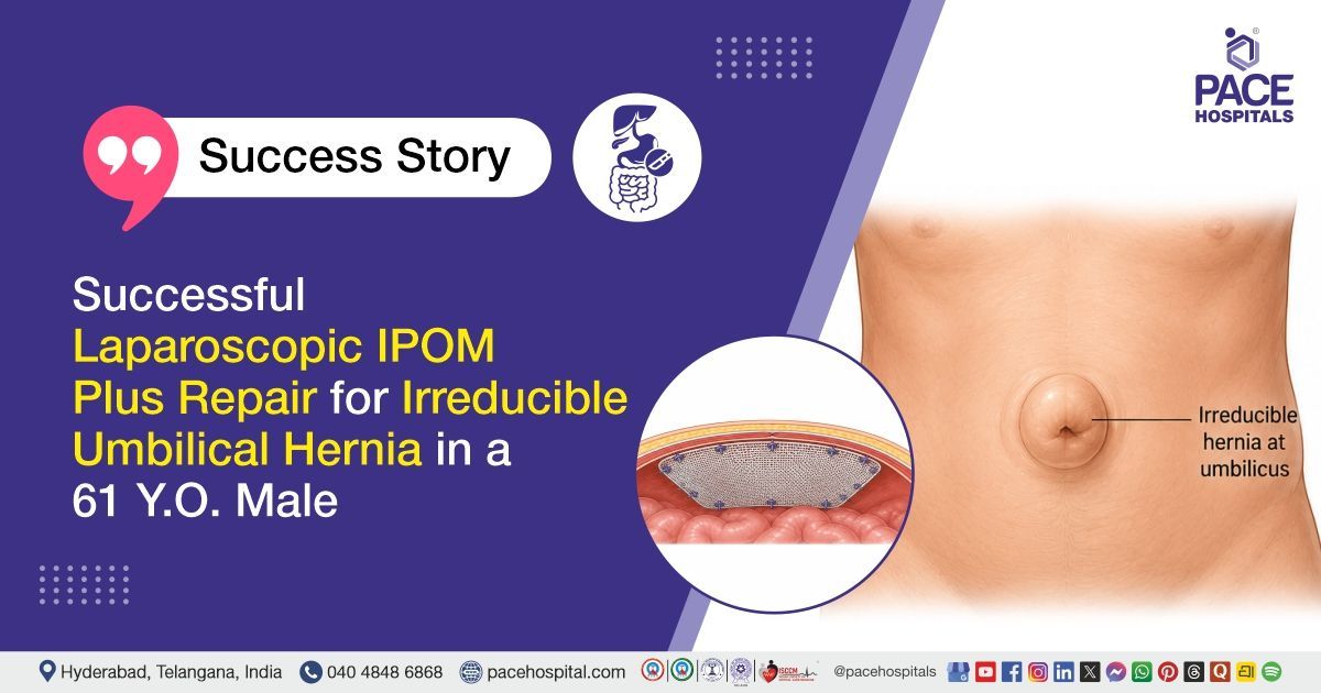
PACE Hospitals’ expert Surgical Gastroenterology team successfully performed a Laparoscopic Intraperitoneal Onlay Mesh (IPOM) Plus Repair on a 61-year-old male patient diagnosed with an irreducible umbilical hernia. The procedure was undertaken to safely reposition the herniated abdominal contents, close the hernia defect, reinforce the weakened abdominal wall with a surgical mesh, relieve symptoms, and reduce the risk of complications and hernia recurrence.
Chief Complaints
A 61-year-old male patient with a body mass index (BMI) of 19 presented to the Surgical Gastroenterology Department at PACE Hospitals, Hitech City, Hyderabad, with a chief complaint of a swelling above the umbilicus (Navel) that had been present for the past two years. The swelling had an insidious onset and progressively increased in size, measuring approximately 5 × 5 cm at the time of presentation. It was irreducible and was not associated with any identifiable aggravating or relieving factors. The patient reported no history of pain, abdominal distension, obstipation, fever, vomiting, or any symptoms suggestive of intestinal obstruction.
Past Medical History
The patient was a known case of hypertension for the past 20 years and was on regular treatment. There was no documented history of diabetes mellitus, thyroid disorders, chronic kidney disease, or any other significant systemic illness.
On Examination
On examination, the patient was conscious, coherent, oriented, and hemodynamically stable. Abdominal examination revealed an irreducible swelling in the umbilical region with intact overlying skin. The swelling was non-tender, with no signs of local inflammation. There was no abdominal distension, guarding, or rigidity, and bowel sounds were present. Examination of the cardiovascular, respiratory, and central nervous systems revealed no significant abnormalities.
Diagnosis
Upon admission to PACE Hospitals, following a detailed clinical assessment, the Surgical Gastroenterology team evaluated the patient for complaints of a progressively enlarging swelling above the umbilicus for two years, which had become irreducible. A detailed review of the patient’s relevant medical history, including longstanding hypertension, was undertaken as part of the preoperative evaluation.
Clinical examination revealed an irreducible umbilical swelling without signs of acute intestinal obstruction or strangulation, consistent with a chronic incarcerated umbilical hernia.
Routine investigations, including complete blood count, serum biochemistry, electrolytes, renal function tests, thyroid profile, glycosylated hemoglobin (HbA1c), and urine analysis with culture and sensitivity, were performed as part of the comprehensive preoperative workup. Ultrasound abdomen revealed a defect in the umbilical region measuring approximately 2.5 cm, with herniation of omentum/mesentery noted through the defect showing cough impulse. These findings supported and confirmed the clinical diagnosis.
Based on the confirmed diagnosis, the patient was advised to undergo Irreducible Umbilical Hernia Treatment in Hyderabad, India, under the expert care of the Surgical Gastroenterology Department.
Medical Decision Making (MDM)
After a detailed consultation with Dr. CH Madhusudhan (Senior Consultant Surgical Gastroenterologist and Liver Transplant Surgeon), and cross consultation with Dr. Abhik Debnath (Consultant Laparoscopic Urologist), a comprehensive clinical evaluation was performed to determine the most appropriate management approach for the patient presenting with an irreducible swelling above the umbilicus of two years' duration. Given the patient's longstanding history of hypertension, a thorough preoperative assessment was undertaken to evaluate his fitness for surgery and optimize perioperative safety.
Considering the patient's history of a progressively enlarging irreducible umbilical swelling without features of acute bowel obstruction or strangulation, along with the clinical and radiological findings of an umbilical hernia containing omental contents, the diagnosis of an irreducible umbilical hernia was confirmed. Routine laboratory investigations and pre-anesthetic evaluation were completed and found to be satisfactory for surgical intervention.
Based on the clinical assessment and preoperative evaluation, it was determined that Laparoscopic Intraperitoneal Onlay Mesh (IPOM) Plus Repair was the most appropriate treatment strategy. The procedure was planned to reduce the herniated omental contents, close the fascial defect, reinforce the abdominal wall with a composite mesh, minimize the risk of recurrence, and provide the benefits of a minimally invasive approach, including reduced postoperative pain, shorter hospital stay, and faster recovery.
The patient and his family members were counselled regarding the diagnosis, the proposed surgical procedure, its benefits and potential risks, expected intraoperative findings, postoperative recovery, and the importance of adherence to postoperative instructions and scheduled follow-up to ensure optimal healing and long-term outcomes.
Surgical Procedure
Following the decision, the patient was scheduled to undergo Laparoscopic Intraperitoneal Onlay Mesh (IPOM) Plus Repair Surgery in Hyderabad at PACE Hospitals, under the expert care of the Surgical Gastroenterology Department.
The procedure involved the following steps:
- Patient Preparation and Port Placement: The patient was placed under general anesthesia in a supine position. After aseptic preparation and draping of the abdomen, pneumoperitoneum was created. Standard laparoscopic ports were inserted under direct vision to access the peritoneal cavity and evaluate the hernia defect.
- Exploration and Adhesiolysis: Intraoperative exploration revealed a 3 × 3 cm umbilical hernia defect containing omental tissue. Adhesiolysis was carefully performed to release intra-abdominal adhesions, and the herniated omentum was reduced back into the peritoneal cavity.
- Hernia Sac Management and Defect Access: The hernia sac was addressed, and excess overlying skin was excised. The fascial defect was then accessed through an open approach to facilitate secure closure. The defect edges were clearly delineated for primary repair.
- Primary Fascial Closure and Mesh Placement: The hernia defect was closed primarily using non-absorbable prolene sutures (1-0), reinforcing the abdominal wall. A 20 × 25 cm composite mesh was then positioned in the intraperitoneal space to provide additional reinforcement, ensuring adequate overlap beyond the defect margins.
- Mesh Fixation and Completion of Procedure: The mesh was secured laparoscopically using absorbable tackers to prevent migration and ensure proper fixation. Hemostasis was confirmed, ports were removed under vision, and port sites were closed in layers. The procedure was completed without complications.
Postoperative Care
The surgical procedure itself was uneventful, and the patient initially had a smooth recovery. On postoperative day 2, the patient developed abdominal bloating, abdominal pain, distension, a fever episode, urinary retention, and burning micturition, for which a urology consultation was obtained, and relevant investigations, including cultures, were sent, followed by appropriate modification of management.
The patient showed gradual clinical improvement with resolution of symptoms, including relief from abdominal pain and restoration of normal bowel function. He improved with supportive medical care aimed at maintaining hydration, controlling infection, and relieving gastrointestinal symptoms and was discharged in a hemodynamically stable condition.
Discharge Medications
Upon discharge, the patient was prescribed medications for prevention and treatment of post-operative infection, gastric protection, pain control, and relief of post-surgical discomfort. Supportive medications were advised to improve bowel function and relieve constipation, along with multivitamin supplementation to support recovery and wound healing.
Treatment for blood pressure control was continued as per existing management. Additional short-term medications were provided for urinary symptom relief and to support hydration during recovery. Medications for cardiovascular risk prevention were also continued as part of ongoing care.
Advice on Discharge
The patient was advised to continue a normal diet as tolerated. Post-operative instructions included the use of an abdominal binder for 8 weeks to support the surgical site and promote healing. The patient was also advised to avoid straining, heavy physical activities, and sitting on the floor to prevent increased intra-abdominal pressure and reduce the risk of recurrence or complications.
Emergency Care
The patient was advised to report to the emergency ward at PACE Hospitals in case of fever, abdominal pain, or vomiting.
Review and Follow-up Notes
The patient was advised to return for a follow-up visit with the Surgical Gastroenterologist in Hyderabad at PACE Hospitals after 3 days.
Conclusion
This case highlights a patient with an irreducible umbilical hernia managed successfully with laparoscopic IPOM Plus repair. The postoperative period was uneventful following appropriate supportive management of transient symptoms, with steady clinical improvement. The patient was discharged in a hemodynamically stable condition with advice for regular follow-up and postoperative precautions.
Abdominal Wall Hernia Management Using Minimally Invasive Laparoscopic Techniques
Minimally invasive approaches have become a preferred standard in the management of abdominal wall hernias due to improved safety profiles and reduced postoperative morbidity. Laparoscopic mesh-based repair techniques enable effective defect closure with strong reinforcement of the abdominal wall, supporting long-term durability and lowering recurrence risk. Careful clinical assessment and appropriate imaging are essential to confirm diagnosis and determine the optimal surgical approach under a Surgical gastroenterologist / Surgical gastroenterology doctor. Intraoperative principles include safe reduction of hernial contents, adhesiolysis when indicated, and precise mesh placement for reinforcement. Postoperative recovery is generally faster, with early mobilization, reduced pain, and quicker return of gastrointestinal function. Overall, multidisciplinary care ensures optimal outcomes and enhanced patient recovery.
Frequently Asked Questions (FAQs)
What is laparoscopic IPOM Plus repair for an irreducible umbilical hernia?
Laparoscopic IPOM Plus repair is a minimally invasive procedure used to treat an umbilical hernia that cannot be pushed back into the abdomen. During the surgery, the hernia contents are returned to their normal position, the abdominal wall defect is stitched closed, and a surgical mesh is placed over it for extra support. This helps strengthen the abdominal wall and lowers the chance of the hernia coming back.
Why is mesh used during umbilical hernia repair?
A surgical mesh is used to reinforce the weakened area of the abdominal wall after closing the hernia defect. It provides additional strength while the tissues heal and significantly reduces the risk of hernia recurrence, especially in larger or long-standing hernias. Modern composite meshes are designed to be safe and well tolerated by the body.
What does it mean if an umbilical hernia is irreducible?
An irreducible umbilical hernia means the bulge cannot be pushed back into the abdomen because the tissues have become trapped. Although it may not always cause severe pain, it has a higher risk of complications such as bowel obstruction or loss of blood supply to the trapped tissue. Surgery is generally recommended to prevent these problems.
How long does recovery take after laparoscopic IPOM Plus hernia repair?
Most patients are able to walk within a day after surgery and return to light daily activities within one to two weeks. Complete healing of the abdominal wall usually takes around six to eight weeks. Recovery may vary depending on the size of the hernia, overall health, and whether any complications occurred after surgery.
Why do doctors recommend wearing an abdominal binder after hernia surgery?
An abdominal binder provides gentle support to the healing abdominal muscles and helps reduce discomfort while moving, coughing, or standing. It also helps patients feel more comfortable during recovery. It should be worn only for the duration advised by the surgeon and should not replace activity restrictions.
Can complications occur after laparoscopic umbilical hernia repair?
Most patients recover well after laparoscopic hernia repair. In some cases, mild bloating, constipation, difficulty passing urine, low-grade fever, or wound discomfort may occur for a short time. These problems usually settle with medicines and proper care. However, severe abdominal pain, repeated vomiting, or high fever should be reported to the doctor immediately.
When can I return to work and normal daily activities after surgery?
People with desk jobs may be able to return to work within one to two weeks, depending on their recovery. Jobs that involve heavy lifting or strenuous physical work usually require a longer recovery period of four to eight weeks. The exact timing should be decided during follow-up visits with the surgeon.
What activities should be avoided after umbilical hernia repair?
Patients should avoid heavy lifting, strenuous exercise, and any activity that puts pressure on the abdomen during recovery. While coughing, the abdomen should be supported gently if advised. Straining during bowel movements and sitting on the floor should also be avoided for some time. These precautions help the repaired area heal well and reduce the chance of hernia recurrence.
Can an umbilical hernia come back after laparoscopic mesh repair?
Although laparoscopic mesh repair has a low recurrence rate, no surgery can completely eliminate the possibility of a hernia returning. The risk is lower when the defect is properly repaired with mesh and patients follow post-operative instructions. Maintaining a healthy weight, avoiding smoking, and preventing chronic constipation or persistent coughing can further reduce the chances of recurrence.
When should I seek immediate medical attention after hernia surgery?
Contact your doctor or go to the emergency department if you have severe abdominal pain, repeated vomiting, high fever, wound redness or discharge, difficulty passing urine, inability to pass stools or gas, or sudden swelling near the surgery area. Early treatment can help prevent serious complications.
Share on
Request an appointment
Fill in the appointment form or call us instantly to book a confirmed appointment with our super specialist at 04048486868
Appointment request - health articles
Recent Articles