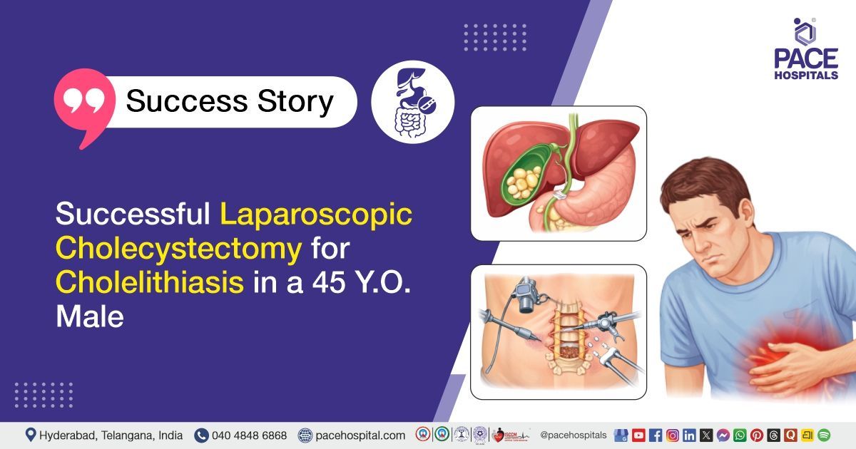
Successful Laparoscopic Cholecystectomy for Cholelithiasis in a 45 Y.O. Male
PACE Hospitals
PACE Hospitals’ expert Surgical Gastroenterology team successfully performed a Laparoscopic Cholecystectomy on a 45-year-old male patient diagnosed with symptomatic cholelithiasis. The aim of the procedure was to safely remove the gallbladder containing stones, relieve symptoms such as abdominal pain and discomfort, and prevent potential complications, including infection, obstruction, or pancreatitis.
Chief Complaints
A 45-year-old male patient with a body mass index (BMI) of 21 presented to the Surgical Gastroenterology Department at PACE Hospitals, Hitech City, Hyderabad, with chief complaints of right upper quadrant abdominal pain of moderate to severe intensity, colicky(coming in waves or cramps) in nature, persisting for the past one week and temporarily relieved with medication. The pain was associated with two episodes of bilious vomiting.
Past Medical History
The patient was a known case of hypertension for one year and was on regular treatment. He had a history of alcoholic hepatitis with liver fibrosis, which had been managed conservatively in the past. He had also experienced two similar episodes of right upper quadrant abdominal pain previously, suggesting symptomatic cholelithiasis. There was no history of diabetes mellitus, coronary artery disease, chronic kidney disease, or any prior major surgeries.
On Examination
On examination, the patient was conscious, coherent, and oriented, and was afebrile and hemodynamically stable. Cardiovascular system examination was normal with normal heart sounds and no murmurs. Respiratory system examination revealed normal bilateral air entry with no added sounds. Abdominal examination showed a soft abdomen with tenderness in the right upper quadrant, without guarding, rigidity, or palpable mass, and bowel sounds were normal. Central nervous system examination was normal.
Diagnosis
Upon admission to PACE Hospitals, the patient was thoroughly evaluated by the Surgical Gastroenterology team. He presented with moderate to severe colicky pain in the right upper quadrant of the abdomen associated with two episodes of bilious vomiting. He also had a history of similar recurrent episodes in the past. Clinical assessment and history were suggestive of symptomatic gallstone disease.
The patient underwent further diagnostic evaluation. Ultrasound abdomen and CT abdomen revealed multiple gallbladder calculi (approximately 30 gallstones, the largest measuring 7 mm)with mild gallbladder wall thickening and features suggestive of mild calculus cholecystitis, along with hepatomegaly and grade I fatty liver changes. Fibroscan indicated mild to moderate liver fibrosis. Routine laboratory investigations, including complete blood picture, liver function tests, renal function tests, coagulation profile, viral markers, and urine examination, were performed. Most parameters were within normal limits, with mild abnormalities noted in coagulation profile and urine examination. Cardiac evaluation, including ECG and 2D echocardiography, showed normal cardiac function.
Based on the confirmed diagnosis, the patient was advised to undergo Gallstones Treatment in Hyderabad, India, under the expert care of the Surgical Gastroenterology Department, to relieve symptoms and prevent further complications.
Medical Decision Making
After a detailed consultation with the consultant Surgical Gastroenterologist, Dr. Suresh Kumar S, a thorough evaluation was conducted considering the patient complaints of moderate to severe colicky right upper quadrant abdominal pain associated with two episodes of bilious vomiting, with a history of two similar previous episodes. All relevant laboratory and imaging investigations including complete blood picture, liver function tests, renal function tests, coagulation profile, viral screening, ultrasound abdomen, CT abdomen, Fibroscan, chest X-ray, ECG, and 2D ECHO were reviewed.
Based on the clinical and imaging investigations, it was determined that laparoscopic cholecystectomy was identified as the most appropriate intervention to remove gallstones, relieve symptoms, and prevent further complications associated with this condition.
The patient and his family members were counselled regarding the diagnosis, the procedure, risks, and its potential to relieve symptoms and enhance the quality of life.
Surgical Procedure
Following the decision, the patient was scheduled to undergo Laparoscopic Cholecystectomy Surgery in Hyderabad at PACE Hospitals under the expert care of the Surgical Gastroenterology Department.
The following steps were done during the procedure:
- Access and Port Placement: The patient was placed in a supine position under general anesthesia. Pneumoperitoneum was created using a Veress needle, and intra-abdominal pressure was maintained at standard levels. Four laparoscopic ports were inserted: one at the umbilicus for the camera, one in the epigastric region, and two in the right upper quadrant for working instruments. Adequate visualization of the gallbladder and Calot’s triangle was ensured.
- Exposure and Adhesiolysis: The gallbladder was found to be distended and multiple stones (approximately 30 gallstones, the largest measuring 7 mm) were noted. Adhesions around the gallbladder and Calot’s triangle were carefully dissected (adhesiolysis) to allow safe exposure of the cystic duct and cystic artery. Fatty tissue in Calot’s triangle was carefully cleared for proper identification of structures.
- Dissection of Calot’s Triangle and CVS Achievement: Meticulous dissection of the Calot’s triangle was performed to clearly identify the cystic duct and cystic artery. The critical view of safety (CVS) was achieved to ensure no injury to the common bile duct or hepatic structures.
- Ligation and Division of Cystic Duct and Artery: The cystic artery was clipped proximally and distally and then divided. Similarly, the cystic duct was clipped on both ends and divided. Care was taken to avoid injury to the gallbladder wall and liver bed during this step.
- Gallbladder Removal and Closure: The gallbladder was dissected from the liver bed using careful electrocautery for hemostasis. Once completely free, the specimen was extracted through the umbilical port. Ports were closed in layers, and the procedure was completed without complications.
Postoperative Care
The procedure was uneventful, and the patient’s postoperative recovery has been satisfactory. During the hospital stay, he was managed for infection prevention, pain control, and gastric protection. He experienced no significant complications and remained stable at the time of discharge.
Discharge Medications
Upon discharge, the patient was prescribed medications to prevent infection, reduce stomach acid, manage nerve-related symptoms, relieve pain as needed, and support gallbladder and liver health.
Dietary Advice
The patient was advised to follow a regular, well-balanced diet to support recovery and maintain overall health.
Emergency Care
The patient was informed to contact the emergency ward at PACE Hospitals in case of any emergency or development of symptoms such as fever, abdominal pain ,Chest pain and vomiting.
Review and Follow-up Notes
The patient was advised to return for a follow-up visit with the Surgical Gastroenterologist in Hyderabad at PACE Hospitals, after 3 days, for further evaluation.
Conclusion
This case highlights symptomatic cholelithiasis managed with laparoscopic cholecystectomy. The procedure was uneventful, and postoperative recovery was smooth without complications. The patient remained hemodynamically stable and was discharged with medications to prevent infection, manage pain, support liver and gallbladder function, and aid recovery through a regular diet.
Integrative Care in Symptomatic Cholelithiasis Management
Managing symptomatic gallstones with mild cholecystitis and fatty liver changes benefits from an integrative approach. A surgical gastroenterologist / surgical gastroenterology doctor performs a laparoscopic cholecystectomy with meticulous attention to Calot’s triangle, ensuring safe and complete stone removal. Coordination with hepatology and primary care optimizes management of hepatic fibrosis and comorbidities. Postoperative care, including targeted nutrition, analgesia, antibiotics, and hepatoprotective medication, promotes rapid recovery. Regular follow-up laboratory tests and imaging allow early detection of complications, demonstrating the importance of combining surgical, medical, and nutritional strategies for optimal outcomes.
Share on
Request an appointment
Fill in the appointment form or call us instantly to book a confirmed appointment with our super specialist at 04048486868
Appointment request - health articles
Recent Articles