Pleural Effusion: Symptoms, Causes & Treatment
PACE Hospitals
Written by: Editorial Team
Medically reviewed by: Dr. Pradeep Kiran Panchadi - Consultant Pulmonologist, Specialist in Bronchoscopy and EBUS
Overview | Incidence | Types | Symptoms | Causes | Risk Factors | Complications | Diagnosis | Treatment | Prevention | Pulmonary edema vs Pleural effusion | Transudative vs Exudative pleural effusion | FAQs | When to consult a Doctor
Pleural effusion definition
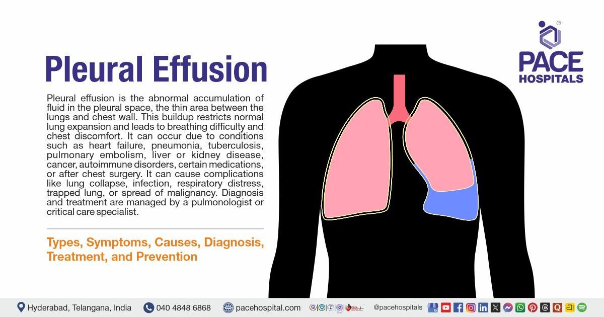
Pleural effusion is an abnormal accumulation of excess fluid between the pleural space (a thin, fluid-filled area between the two layers of the pleura, which are membranes that surround the lungs), causing breathing difficulties and chest discomfort. Common symptoms may include dyspnea (shortness of breath), chest pain (often sharp and worsens with deep breaths), dry cough, low-grade fever, and, in some cases, reduced exercise tolerance or fatigue.
The main causes include heart failure, pneumonia, tuberculosis (TB), pulmonary embolism, liver disease (cirrhosis), kidney disease, cancer (lung, breast, lymphoma, metastatic disease), autoimmune conditions (such as lupus or rheumatoid arthritis (RA)), certain medications, and post-thoracic surgery. If left untreated, pleural effusion can lead to major complications such as respiratory distress, lung collapse (atelectasis), recurrent infections (empyema), trapped lung (fibrosis preventing lung expansion), and, in severe cases, sepsis or spread of malignancy within the pleura.
A pulmonologist, a specialist in lung diseases, and a critical care physician, who specializes in managing patients with life-threatening conditions, can accurately diagnose pleural effusion and recommend a treatment plan based on its underlying cause.
Pleural effusion meaning
The term "pleural effusion" is derived from a combination of Greek and Latin roots.
The word's origins can be broken down as follows:
- "pleura": which refers to the side, rib, or membrane surrounding the lungs.
- "effundo": which means "shed," "pour forth," or "pour out"
Together, pleural effusion literally means “a pouring out into the pleural space”, describing the abnormal accumulation of fluid between the layers of the pleura around the lungs.
Incidence of Pleural Effusion
Incidence of pleural effusion Worldwide
Pleural effusion is a frequent pleural disease worldwide, with an estimated annual occurrence of 15 lakh cases in the United States and a prevalence of about 320 cases per 100,000 people in industrialised countries. Most cases are caused by heart failure, pneumonia, cancer, and tuberculosis, and rates vary depending on regional disease patterns and healthcare access.
Incidence of pleural effusion in India
In India, tuberculosis (TB) is the leading cause of pleural effusion, with 44–80% of cases reported in recent hospital-based studies, with malignancy comprising the second most common etiology at 13–18%. The disease predominantly affects males and middle-aged adults, with a higher occurrence in resource-limited and infection-prone regions. Seasonal peaks are observed during rainy and winter months, with incidence rates among hospitalised patients ranging from about 2–3% of admissions and higher in children and those from lower socioeconomic backgrounds.

Types of Pleural Effusion
Pleural effusions can be categorised in multiple ways based on the nature and content of the pleural fluid and underlying pathology. The pleural effusion classification is as follows:
- Based on the nature of the fluid (by pathophysiology)
- Transudative pleural effusion
- Exudative pleural effusion
- Based on fluid content
- Serous fluid (hydrothorax)
- Blood (haemothorax)
- Chyle (chylothorax)
- Pus (pyothorax or empyema pleural effusion)
- Urine (urinothorax pleural effusion)
- Specific types of exudative effusions
- Parapneumonic pleural effusion
- Uncomplicated parapneumonic effusions
- Complicated parapneumonic effusions
- Empyema thoracis
- Malignant pleural effusion
- Tuberculous pleural effusion
Based on the nature of the fluid
Transudative pleural effusion
Transudative effusion occurs when there is an imbalance between the pressures controlling fluid movement across the pleura, specifically, increased hydrostatic pressure or decreased oncotic pressure. This causes leakage of a clear, watery fluid that is low in protein and cells. The pleura remains intact and non-inflamed. Common causes include congestive heart failure, liver cirrhosis, and nephrotic syndrome. This leads to the leakage of a clear, watery fluid low in protein and cells.
Exudative pleural effusion
Exudative pleural effusion develops when inflammation or injury to the pleural membrane increases vascular permeability, allowing proteins, enzymes, and immune cells to leak into the pleural space. The fluid is usually cloudy, rich in protein, and may contain inflammatory cells. It commonly occurs in pneumonia (parapneumonic effusion), tuberculosis, malignancy, or autoimmune diseases such as lupus.
Based on fluid content
Hydrothorax
This involves the collection of serous (watery) fluid, typically due to conditions like heart failure, cirrhosis, or nephrotic syndrome. The fluid is clear and transudative in nature.
Hemothorax
It refers to the accumulation of blood in the pleural space, most commonly caused by trauma, ruptured blood vessels, or malignancy. It presents as bloody fluid and is a medical emergency requiring immediate drainage.
Chylothorax
This occurs when lymphatic fluid (chyle) leaks into the pleural cavity because of obstruction or damage to the thoracic duct, often due to trauma, lymphoma, or surgical injury. The fluid appears milky white, rich in triglycerides and lymphocytes.
Empyema pleural effusion
This results from bacterial infection of the pleural space, leading to the accumulation of thick, purulent pus. It often follows pneumonia or lung abscess and needs prompt drainage and antibiotic treatment to prevent fibrosis or sepsis.
Urinothorax
This is a rare type of pleural effusion that occurs when urine leaks into the pleural space, usually following urinary tract obstruction or trauma. The fluid is pale yellow, has a distinct ammonia-like odour, and generally shows a low pH and high creatinine level compared to serum. It resolves once the underlying urinary issue is corrected.
Specific types of exudative effusions
Parapneumonic pleural effusion
This is the pleural fluid accumulations that occur as a complication of bacterial pneumonia, lung abscess, or bronchiectasis, and is classified into three subtypes based on the characteristics of the pleural fluid and the stage of the disease.
- Uncomplicated parapneumonic effusion: This subtype arises early in pneumonia when sterile inflammatory fluid accumulates due to increased capillary permeability. It does not contain bacteria and usually resolves completely with appropriate antibiotic treatment alone without the need for drainage.
- Complicated parapneumonic effusion: If bacterial invasion of the pleural space occurs, the effusion becomes complicated and the pleural fluid turns cloudy. Even though bacteria may sometimes not be detected, the inflammatory process is significant enough to require chest tube drainage in addition to antibiotics for resolution.
- Empyema thoracis: This is the most severe stage, characterised by the presence of frank pus in the pleural cavity, and positive bacterial cultures or Gram stains. The pleural cavity may develop thick loculations and pleural thickening, leading to lung restriction if untreated. Management requires prompt and aggressive drainage, intravenous antibiotics, and sometimes surgical management.
Malignant pleural effusion
This develops when cancer cells invade the pleura or obstruct lymphatic flow, resulting in a pleural fluid that is high in proteins. Common cancer causing these effusions are lung, breast, and mesothelioma (a rare and aggressive cancer of the mesothelium). These effusions generally indicate advanced disease and require ongoing management for symptom relief.
Tuberculous pleural effusion
This is caused by infection of the pleura by Mycobacterium tuberculosis; this exudate usually develops in regions where tuberculosis is prevalent. It is characterised by immune-mediated inflammation with a lymphocyte-predominant fluid and has distinct diagnostic and treatment considerations.

Pleural Effusion Symptoms
The symptoms of pleural effusion may vary based on the amount of fluid deposited and the underlying cause. Recognising these signs of pleural effusion helps in early diagnosis and management. The most common pleural effusion signs and symptoms are as follows:
Common symptoms are:
- Chest pain
- Cough
- Shortness of breath (SOB)
- Fever
Less common symptoms are:
- Weakness
- Hiccups
- Rapid breathing
- Weight loss
Chest pain: This symptom is sudden and gets worse with coughing, deep breathing, or movement. It is caused by inflammation of the pleural lining (pleurisy), in which the two layers of pleura rub against each other, resulting in pain. The pain tends to decrease if enough fluid accumulates to separate these layers and prevent friction.
Cough: Coughs are usually a dry, non-productive cough that arises due to irritation of the lung or pleura adjacent to the effusion. It is not caused by mucus but by the mechanical effect of fluid and inflammation.
Shortness of breath (SOB): Shortness of breath is the most common symptom and occurs because the fluid compresses the lung, limiting its expansion and reducing effective lung volume. The difficulty breathing may worsen with exertion and increase as fluid volume grows.
Fever: Fever occurs mainly in exudative effusions caused by infection, such as pneumonia or tuberculosis. It results from the body’s immune response to inflammation or infection within the pleura.
Weakness: Generalised weakness as well as fatigue are caused by a lack of oxygen, systemic inflammation, and the body's effort to fight infection or cancer. Chronic effusions can also cause energy depletion and muscle loss.
Hiccups: The irritation or compression of the diaphragm by accumulated fluid can stimulate the phrenic nerve, resulting in persistent hiccups. This is an uncommon but characteristic symptom in large effusions.
Rapid breathing (tachypnoea): Reduced lung capacity and oxygen levels cause the body to compensate by increasing the breathing rate. This helps maintain an adequate oxygen supply but can cause discomfort and fatigue.
Weight loss: Weight loss is particularly seen in chronic or disease-related effusions, such as tuberculosis or cancer. It results from prolonged illness, poor appetite, and the body’s increased metabolic demand during chronic inflammation or malignancy.

Pleural Effusion Causes
Pleural effusion occurs when the normal balance between pleural fluid production and absorption is disturbed. This imbalance can result from systemic conditions or local disease processes. The causes are generally classified by Light's criteria for pleural effusion based on whether the effusion is transudative or exudative.
The most common causes of pleural effusion are:
Transudative causes
- Congestive heart failure
- Cirrhosis
- Kidney disease
- Pulmonary embolism
Exudative causes
- Infections
- Cancer
- Autoimmune diseases
- Pancreatitis
- Post-cardiac surgery
- Trauma
- Certain drugs
- Radiotherapy
Transudative Causes
These occur due to systemic factors that alter hydrostatic or oncotic pressure, leading to fluid leakage without inflammation of the pleura, which include:
Congestive heart failure (CHF): This is the most common cause of pleural effusion. When the heart can't pump blood efficiently, pressure builds up in the pulmonary circulation. The increased hydrostatic pressure forces fluid out of the blood vessels into the pleural space. The fluid is usually clear and low in protein (transudate). Effusions are bilateral and will resolve with adequate heart failure care.
Cirrhosis: In advanced liver disease, especially cirrhosis, portal hypertension and low blood protein levels (hypoalbuminemia) cause fluid to leak from the abdominal cavity into the pleural space through small openings in the diaphragm. This type, known as hepatic hydrothorax, usually presents as a right-sided pleural effusion and is transudative in nature.
Kidney disease: Conditions like nephrotic syndrome or chronic kidney failure cause loss of protein in urine and low blood albumin levels. This reduces the plasma oncotic pressure, allowing fluid to move into the pleural cavity. In severe renal failure, excess body fluid accumulation (fluid overload) may also contribute to pleural effusion formation.
Pulmonary embolism: A blood clot in the lung artery can increase pressure in the lung circulation, leading to transudative effusion. However, if the embolism causes inflammation or tissue injury, it may also result in an exudative effusion. Symptoms often include sudden shortness of breath and chest pain.
Exudative Causes
These result from local factors, such as inflammation, infection, or malignancy, which increase vascular permeability or impair lymphatic drainage, including:
Infections: Infection from bacteria, viruses, or fungi can inflame the pleura, causing fluid to leak into the pleural cavity. Pneumonia can lead to parapneumonic effusion, and if the infection spreads, it can progress to empyema, where pus accumulates in the pleural space.
Cancer: Malignant tumours, including lung cancer, breast cancer, and lymphomas, can penetrate or irritate the pleural membrane, increase capillary permeability, or block lymphatic outflow. This leads to the accumulation of exudative, usually blood-stained fluid. Malignant pleural effusions tend to be recurring and indicate severe disease.
Autoimmune diseases: Certain autoimmune diseases, such as Rheumatoid arthritis (RA) and lupus, can cause pleural inflammation (pleuritis), leading to exudative effusion. The pleural effusion fluid generally contains high levels of inflammatory cells and proteins. Patients may also notice joint pain, fatigue, or rash due to the underlying systemic condition. Rheumatoid arthritis and lupus commonly cause bilateral pleural effusion.
Pancreatitis: In acute or chronic pancreatitis, digestive enzymes like amylase can travel through the diaphragm or bloodstream and irritate the pleura, resulting in exudative pleural effusion. The fluid often contains high levels of amylase and is usually associated with left pleural effusion.
Post-cardiac surgery: After heart surgery, inflammation and irritation of the pleura or pericardium can lead to fluid accumulation in the pleural space. This exudative effusion is often sterile and resolves gradually, though persistent effusions may require drainage.
Trauma (post-traumatic pleural effusion): Injury to the chest can rupture blood vessels or lymphatic channels, causing a build-up of blood (hemothorax) or lymph (chylothorax) in the pleural space. Trauma-related effusions are exudative and may require surgical intervention to prevent complications like infection or lung compression.
Certain drugs: Certain drugs, such as disease-modifying antirheumatic drugs (DMARDs), antiarrhythmic agents, anticonvulsants (hydantoin derivatives), and tyrosine kinase inhibitors (TKIs), can induce pleural inflammation or lupus-like reactions, leading to exudative pleural effusion. Drug-induced effusions usually resolve after stopping the offending medication.
Radiotherapy: Radiation therapy to the chest for cancer can cause delayed inflammation, scarring, or damage to pleural capillaries and lymphatic vessels. This results in exudative pleural effusion, which may appear months after treatment.

Pleural Effusion Risk Factors
Pleural effusion arises when underlying medical disorders or risk factors disrupt the balance of fluid production and absorption in the pleural space. They are:
Non-controllable risk factors
- Age
- Gender
- Family history
Controllable risk factors
- Pre-existing medical conditions
- Lifestyle habits
- Medicinal history
- Occupational exposure to toxins
Non-controllable risk factors
Age: The risk of certain causes, such as heart disease and cancer, increases with age. Some sources note a higher risk for certain pleural disorders in older adults and specific age ranges.
Gender: Men are more prone to develop effusions from pneumonia, heart disease, and occupational exposures, whereas women are more likely to develop effusions linked to autoimmune diseases like lupus or RA. Hormonal differences and lifestyle factors can also influence susceptibility.
Family history: A gene of predisposition to conditions like heart disease, kidney disorders, or autoimmune diseases can indirectly increase the risk of pleural effusion. Family history of tuberculosis or cancer may also raise the likelihood of developing pleural fluid accumulation due to hereditary or environmental factors shared within families.
Controllable risk factors
Pre-existing medical conditions: Chronic diseases such as heart failure, liver cirrhosis, kidney disease, tuberculosis (TB), and cancer may increase the risk of pleural effusion. These diseases either cause fluid overload, reduced protein levels, or direct pleural inflammation, leading to transudative or exudative effusions. Individuals with respiratory infections or pulmonary embolism are vulnerable.
Lifestyle habits: Unhealthy habits such as smoking, chronic alcohol consumption, and poor diet can indirectly raise the risk of pleural effusion. Smoking can damage the lungs and increase susceptibility to infections and lung cancer, while alcohol misuse contributes to liver disease (cirrhosis), a major cause of transudative effusions. A sedentary lifestyle increases the risk of blood clots, which can cause pulmonary embolism and effusion.
Medicinal history: Certain medicines, such as disease-modifying antirheumatic drugs (DMARDs), antiarrhythmic agents, anticonvulsants, and tyrosine kinase inhibitors, can cause drug-induced pleural inflammation or lupus-like reactions, resulting in exudative pleural effusions.
Occupational exposure to toxins: Prolonged exposure to harmful substances, such as asbestos, silica, or industrial chemicals, increases the chance of developing pleural effusion by destroying lung tissue and membranes. Asbestos exposure, in particular, can lead to pleural thickening, effusions, and even mesothelioma (pleural cancer). Workers in mining, construction, chemical industries, or welding are especially at risk.
Pleural Effusion Complications
When pleural effusion is not diagnosed and treated properly, it can lead to complications, including difficulty breathing, infection, and permanent lung damage. The severity of complications may depend on the underlying cause, amount of fluid, and duration of the effusion. Below are the complications of pleural effusion:
- Empyema
- Pleural thickening
- Respiratory failure
- Pneumothorax
- Pleural fibrosis
- Recurrent pleural effusion
Empyema: Empyema is a serious complication that occurs when the fluid in the pleural space becomes infected, turning into thick pus. It often develops as a result of bacterial pneumonia or lung abscess spreading to the pleural cavity. The infected fluid increases inflammation, causes high fever, chest pain, and foul-smelling discharge. If untreated, empyema can form loculated pockets and lead to sepsis or lung scarring.
Pleural thickening: Chronic inflammation or infection of the pleura can cause scarring and thickening of the pleural layers. This pleural thickening reduces the elasticity of the pleural membranes, making it difficult for the lungs to expand fully during breathing. As a result, the person may have symptoms of persistent shortness of breath and chest tightness.
Respiratory failure: When a large amount of pleural fluid compresses the lungs, it limits their ability to expand and exchange oxygen efficiently. This can cause hypoxia (low amounts of oxygen in the blood) and, in severe cases, respiratory failure. The risk increases if both lungs are affected or if the underlying condition, such as heart failure or infection, worsens.
Pneumothorax: Pneumothorax may occur as a complication during pleural fluid drainage procedures like thoracentesis if the lung is accidentally punctured, causing lung collapse and sudden respiratory distress.
Pleural fibrosis: Persistent inflammation or infection can result in the formation of fibrous tissue in the pleural space. It can restrict the lung, preventing it from fully expanding, resulting in long-term breathing problems.
Recurrent pleural effusion: Pleural effusion often comes back even after treatment in some patients. This happens particularly when pleural effusion is caused by long-term conditions such as tuberculosis, cancer, or heart failure. Recurrent effusions cause repeated episodes of shortness of breath, discomfort, and frequent drainage procedures.
Pleural Effusion Diagnosis
Diagnosis of pleural effusion involves a systematic, step-by-step approach to identify the presence of fluid in the pleural space, determine its cause, and guide appropriate treatment. The process combines clinical evaluation, imaging studies, and laboratory analysis of pleural fluid.
The following are the different steps involved in the pleural effusion diagnosis:
- Medical history
- Physical examination
- Imaging studies
- Chest radiograph
- Thoracic ultrasound
- Chest computed tomography (CT scan)
- Echocardiogram
- Thoracentesis (pleural fluid aspiration)
- Pleural fluid analysis
- Light’s criteria (with serum protein, serum LDH)
- pH level
- Pleural fluid glucose level
- Fluid cell count differential
- Fluid gram stain and culture
- Cytological testing
- Laboratory tests
- Complete blood count with differential
- Serum electrolytes
- Serum urea and creatinine
- Liver function tests
- Condition-specific markers
- Acid-fast bacilli smear
- Mycobacterium tuberculosis culture
- NT-proBNP (B-type natriuretic peptide)
- Adenosine deaminase (ADA)
- Amylase
- Pleural fluid triglycerides
- Pleural fluid haematocrit
- Additional/invasive diagnostic tests
- Pleural biopsy
- Medical thoracoscopy (pleuroscopy)
Pleural Effusion Treatment
The goals of treating pleural effusion are to eliminate the accumulated fluid, alleviate symptoms, and address the underlying cause to prevent recurrence. Pleural effusion management is generally determined by whether it is transudative or exudative, size, and the patient's medical state.
Treatment is broadly divided into:
Non-pharmacological management
- Observation
- Supportive care
- Oxygen therapy
- Nutritional support
- Lifestyle modifications
- Breathing exercise and physiotherapy
Medical management of pleural effusion
- For transudative effusions (management of underlying cause)
- Diuretics
- ACE inhibitors or beta-blockers
- Albumin infusion
- For exudative effusion (management of underlying cause)
- Antibiotics
- Corticosteroids
- Immunosuppressants
- Antitubercular drugs
- Chemotherapy/ targeted therapy
Surgical and interventional management
- Therapeutic thoracentesis (pleural fluid drainage)
- Chest tube drainage (intercostal tube)
- Intrapleural fibrinolysis agents
- Pleurodesis (sclerosing agents)
- Indwelling pleural catheter (IPC)
- Video-assisted thoracoscopic surgery (VATS)
- Decortication(for empyema/fibrothorax)
- Medical thoracoscopy (pleuroscopy)
- Pleuroperitoneal shunt
- Thoracic duct ligation (for chylothorax)
Why Choose PACE Hospitals?
Expert Super Specialist Doctors
Advanced Diagnostics & Treatment
Affordable & Transparent Care
24x7 Emergency & ICU Support

Pleural Effusion Prevention
Preventing pleural effusion mainly involves addressing and controlling the underlying diseases or conditions that cause fluid buildup. While not all cases are preventable, the following measures can reduce the risk of developing pleural effusion:
- Promptly treating underlying diseases
- Preventing and controlling infections
- Avoiding exposure to toxins and irritants
- Maintaining healthy lifestyle habits
- Ensuring vaccinations
- Monitoring chronic conditions
- Ensuring post-surgical and hospital care
Promptly treating underlying diseases: Early and effective management of conditions such as heart failure, liver cirrhosis, kidney disease, and cancer helps prevent fluid buildup in the pleural cavity. By controlling over this primary disease, the chances of secondary pleural effusion are greatly reduced.
Preventing and controlling infections: Timely treatment of respiratory infections like pneumonia or tuberculosis prevents inflammation of the pleura. This reduces the risk of fluid accumulation caused by infection-related irritation or damage to the pleural membranes.
Avoiding exposure to toxins and irritants: Avoiding exposure to toxins and irritants such as asbestos or smoking prevents pleural inflammation and malignant transformations that can cause recurrent effusions.
Maintaining healthy lifestyle habits: Healthy habits like regular exercise, balanced nutrition, quitting smoking, and limiting alcohol consumption strengthen the immune and cardiovascular systems. This lowers the likelihood of diseases that can cause pleural effusion, such as heart or lung conditions.
Ensuring vaccinations: Vaccinations against infections such as influenza and pneumococcal pneumonia reduce the risk of respiratory diseases that can lead to pleural inflammation and fluid buildup.
Monitoring chronic conditions: Regular medical check-ups help in the early detection of fluid retention or worsening of chronic diseases such as heart or kidney failure. Early intervention can prevent pleural fluid accumulation before it becomes severe.
Ensuring careful post-surgical and hospital care: This minimises procedure-related infections and trauma, common contributors to effusions after cardiac or thoracic surgery.
Difference between Pulmonary Edema and Pleural Effusion
Both conditions involves fluid accumulation in or around the lungs, but they differ significantly in their location, causes, clinical features, and management. Understanding these differences is crucial for correct diagnosis and targeted treatment. Below are some of the parameters that help in differentiating pulmonary edema and pleural effusion:
Pulmonary edema vs pleural effusion
| Parameters | Pulmonary edema | Pleural effusion |
|---|---|---|
| Definition | Pulmonary edema is the accumulation of fluid within the lung tissues and alveoli (air sacs) that interferes with normal gas exchange. | Pleural effusion is the abnormal collection of fluid in the pleural space, the area between the lungs and the chest wall. |
| Location of fluid | Fluid is present inside the lungs (in the alveoli and interstitial spaces). | Fluid is present outside the lungs (in the pleural cavity). |
| Main causes | Commonly caused by left-sided heart failure, acute respiratory distress syndrome (ARDS), kidney failure, or fluid overload. | Caused by heart failure, infection (pneumonia, tuberculosis), cancer, liver disease, or trauma. |
| Imaging findings | Chest X-ray shows bilateral hazy infiltrates or “bat-wing” appearance due to alveolar fluid. | Chest X-ray or ultrasound shows fluid accumulation at the lung base with blunting of the costophrenic angle. |
| Treatment | Focuses on removing fluid from the lungs—diuretics, oxygen therapy, treating heart failure or the underlying cause. | Focuses on draining pleural fluid (thoracentesis) and treating the underlying cause, such as infection or cancer. |
Difference between Transudate and Exudate Pleural Effusion
Pleural effusion, the abnormal accumulation of fluid in the pleural space, can be broadly classified into transudative and exudative types. This distinction is essential, as it helps narrow down the underlying cause and guides further investigation and treatment. Below are some of the parameters that help in differentiating transudate and exudate pleural effusion:
Transudative vs exudative pleural effusion
| Parameters | Transudative pleural effusion | Exudative pleural effusion |
|---|---|---|
| Definition | Caused by systemic factors that alter pressure or fluid balance across the pleural membranes without inflammation. | Caused by local factors such as inflammation, infection, or malignancy that increase capillary permeability. |
| Main mechanism | Due to increased hydrostatic pressure or decreased oncotic pressure. | Due to increased vascular permeability or impaired lymphatic drainage. |
| Response to treatment | Resolves with treatment of the underlying systemic disorder. | Requires treatment of the local disease and may need drainage or antibiotics. |
Frequently Asked Questions (FAQs) on Pleural Effusion
Can pleural effusion be cured?
Pleural effusion is a condition where fluid builds up around the lungs, and in many cases it can be treated or even cured, depending on the underlying cause. If it is due to infections like tuberculosis or pneumonia, proper medical treatment can completely resolve it. When it is caused by conditions such as heart failure, managing the main disease often reduces the fluid. However, if it is related to cancer, it may not always be fully curable but can still be managed.
Is pleural effusion dangerous?
Pleural effusion can be dangerous depending on its cause and size. Large amounts of fluid can make it difficult to breathe and reduce oxygen delivery. Some causes, like infections or cancer, carry serious risks. If left untreated, complications like lung infection (empyema) or lung collapse can occur. Prompt diagnosis and treatment reduce risks.
How to drain pleural effusion?
Pleural effusion is drained through a procedure called thoracentesis. A doctor inserts a thin needle or tube into the chest wall to remove the excess fluid from the pleural space. This procedure is done with local anaesthesia and ultrasound guidance to ensure safety. In more severe or repeated cases, a chest tube may be inserted for continuous drainage. This helps relieve breathing difficulty and allows laboratory testing of the fluid to find the cause.
Is pleural effusion painful?
Pleural effusion is sometimes painful, but not always. The pain usually depends on whether the lining around the lungs called the pleura is inflamed or not. When pain is present, it is often a sharp or stabbing chest pain that gets worse when taking a deep breath, coughing, or moving, this is called pleuritic pain. However, many people with pleural effusion feel no pain at all and instead notice symptoms like shortness of breath, chest heaviness, or a dry cough.
How is pleural effusion detected in patients?
Doctors detect pleural effusion by listening to the chest with a stethoscope for abnormal sounds and by chest X-rays or ultrasound imaging that show fluid around the lungs. If needed, a needle may be used to withdraw fluid for testing. Advanced imaging, like CT scans or a thoracoscopy (a small camera in the chest), may be done for detailed assessment.
How long does pleural effusion last?
The duration varies widely and depends on the cause and treatment. Effusions from infections may clear with antibiotics in days to weeks. Effusions caused by heart or kidney problems may persist until those conditions are controlled. Some effusions recur or become chronic if the underlying cause is unresolved.
What are the available treatment options for pleural effusion?
Treatment of pleural effusion focuses first on the cause of the extra fluid. For example, if an infection causes it, antibiotics are given. If the fluid causes breathing difficulty, doctors may remove it using a needle or chest tube. In cases of repeated fluid buildup, procedures to prevent fluid return, like pleurodesis (sealing the space), or inserting a permanent drainage catheter, may be done. Surgery is reserved for complicated cases or when the lung can’t fully expand.
Can pleural effusion cause death?
Pleural effusion can become serious and, in some cases, life-threatening, especially if a large amount of fluid compresses the lungs and makes breathing difficult or if the underlying disease is severe. Without timely treatment, complications can increase the risk of death, but many cases are manageable when diagnosed early and treated properly.
What are the causes of left-sided pleural effusion?
Left-sided pleural effusion can occur due to several conditions such as infections (like pneumonia), heart failure, kidney or liver problems leading to fluid buildup, cancer, or blockage of lymphatic drainage. Sometimes, urine leaks from kidney problems can cause fluid to accumulate on the left side. The cause often relates to diseases affecting organs near the left lung or systemic issues that cause fluid to gather in the chest.
Describe the pathophysiology of pleural effusion?
Pleural effusion develops when there is an imbalance between fluid production and absorption in the pleural space. Normally, this space has a small amount of lubricating fluid. When factors such as increased capillary pressure, inflammation, or lymphatic blockage occur, fluid accumulates. This buildup can be transudate (from pressure changes, e.g., heart failure) or exudate (from inflammation or infection), leading to lung compression and breathing difficulty.
What is a bilateral pleural effusion?
A bilateral pleural effusion means that fluid is present on both sides of the chest, surrounding both lungs. It often occurs with diseases that affect the entire body, such as heart failure, kidney or liver disease, or infections. This condition can cause more breathing difficulty than when only one side is affected because both lungs may be compressed.
Can pleural effusion be cured by antibiotics?
Yes, antibiotics can cure pleural effusions caused by infections like pneumonia. However, pleural effusions from other causes, such as heart failure, cancer, or liver disease, require different treatments. Sometimes antibiotics are part of the treatment if infection is involved, but antibiotics alone do not fix effusions caused by other diseases.
How to see pleural effusion on ultrasound?
Ultrasound detects pleural effusion as a dark (fluid) area between the lung and chest wall. The lung appears as moving bright lines (lung sliding). Fluid collects in dependent areas and can be seen as a space where lung movement is reduced. Ultrasound helps measure the fluid amount and guides safe needle or tube placement.
Why right-sided pleural effusion is more common?
Right-sided pleural effusion is often more common because of differences in anatomy and drainage patterns of the lymphatic system. The right lung has a larger surface area, and the right pleural space can more easily collect fluid from liver-related causes such as cirrhosis or hepatic hydrothorax.
When is pleural effusion required to be tapped?
Tapping or thoracentesis is done when pleural effusion causes symptoms like breathlessness or to find out the cause. It is especially done if infection, cancer, or unexplained effusions are suspected. If the effusion is small and not causing problems, tapping may not be needed.
What is the difference between pleural effusion and empyema?
- Pleural effusion is the buildup of fluid in the pleural space, which can result from various conditions such as heart failure, liver cirrhosis, infections, or malignancy.
- Empyema is a type of pleural effusion where the pleural fluid becomes infected and turns into pus. It most commonly occurs as a complication of untreated pneumonia or chest infections.
When to consult a doctor for pleural effusion?
Consult a doctor for pleural effusion if there is shortness of breath, chest pain, or persistent coughing that does not improve with rest. Signs that indicate the need for medical assistance are:
- Difficulty breathing when lying down
- Sharp or pressure-like chest pain during breathing
- Persistent fever or unexplained fatigue
- Swelling in the legs or abdomen
- Cough producing blood or frothy sputum
If these symptoms persist, it is important to visit a pleural effusion doctor for proper diagnosis and treatment. Seek emergency medical help if breathing becomes severe or sudden, as this could indicate a large or infected pleural effusion. A pulmonologist can recommend tests and the best pleural effusion treatment to relieve symptoms and prevent complications.
Share on
Request an appointment
Fill in the appointment form or call us instantly to book a confirmed appointment with our super specialist at 04048486868
Appointment request - health articles
Recent Articles