EBUS Procedure Cost in Hyderabad, India – 2026 Guide
Get a Cost Estimate for EBUS Procedure
Procedure & Surgery Cost Pages
All You Need To Know About the EBUS Procedure
The Endobronchial Ultrasound (EBUS) Procedure is an image-guided, minimally invasive bronchoscopy used to diagnose and stage lung and chest lymph node diseases, without resorting to open surgery.
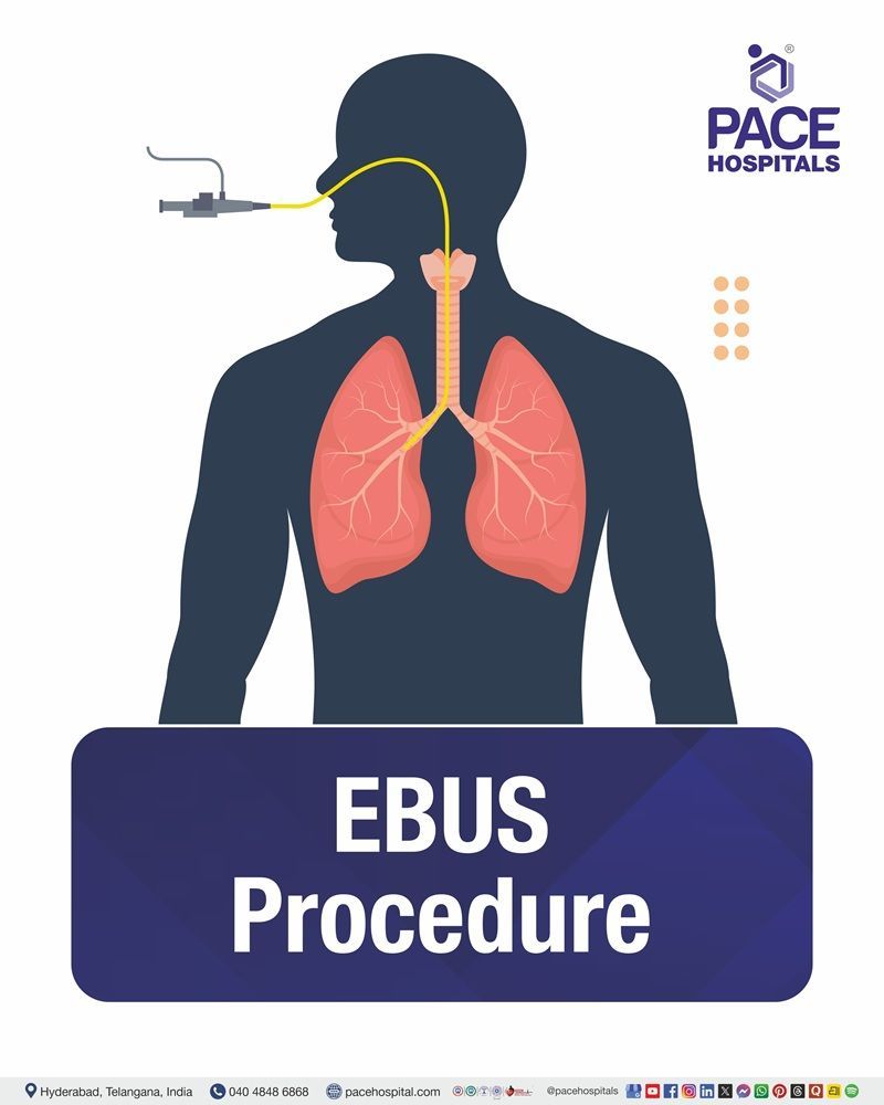
In the EBUS procedure, a thin, flexible bronchoscope fitted with an ultrasound probe is passed through the mouth and into the airways to produce live ultrasound images of the lungs and the surrounding lymph nodes.
During the procedure, the physician can perform Endobronchial Ultrasound-Guided Transbronchial Needle Aspiration (EBUS-TBNA) to collect tissue or lymph node specimens for analysis.
The EBUS Procedure is used to diagnose and stage lung cancer, evaluate enlarged mediastinal/hilar lymph nodes, detect tuberculosis infections, and diagnose sarcoidosis and lymphomas.
This procedure is carried out either under general or moderate sedation, in a day care procedure setting, where patients experience faster recovery and have reduced pain and complications compared with traditional surgical biopsy methods.
EBUS Procedure Cost in India
On average, the cost of an EBUS Procedure in India ranges from ₹18,000 to ₹45,000 and above (approximately US $188 to US $470). However, the actual cost may vary depending on several factors, including:
- Reasons behind the EBUS technique (diagnosis of lung cancer, swollen lymph nodes, infections or any other diseases in the chest)
- Complexity of the EBUS procedure (EBUS-TBNA or EBUS with needle biopsy)
- Number and site of lymph nodes or lung lesions to be sampled
- Age, general health status and other medical conditions of the patient
- Type of hospitals (private, corporate or trust hospitals)
- Costs at the hospital (including room and facility costs)
- Anesthesia/sedation involved in the EBUS procedure
- Experience of pulmonologists and other medical staff
- Pre-procedure investigations (like blood tests, ECG, CT scan of chest or other tests) and post-procedure care requirements
- Insurance or corporate approval for treatment on a cashless basis
EBUS Procedure Cost in Hyderabad, Telangana
On average, the cost of an EBUS procedure in Hyderabad ranges from ₹20,000 to ₹35,000 and above (approximately US $209 to US $366). These rates offer a competitive and cost effective option compared to other top hospitals in the city, with advanced surgical care and personalized treatment planning readily available.
However, the cost of an EBUS Procedure in Hyderabad is influenced by several factors, such as:
- Type of EBUS procedure conducted (EBUS-TBNA or diagnostic bronchoscopy along with EBUS)
- Complexity of the disease and number of lymph nodes/lesions to be sampled
- Infrastructure at the hospital and bronchoscopy lab
- Experience of the interventional pulmonologist and multidisciplinary team participation
- Type of anesthesia or sedation needed for the procedure
- Investigations before procedure, pathology, and laboratory testing of biopsy specimens
- Necessity for further diagnosis using other tests at the same time (such as bronchoscopy/BAL)
- Insurance coverage or corporate approval for cashless treatment
- Post-procedure observation, recovery requirements, and management of any procedure-related complications
Cost Breakdown According to Type of EBUS Procedure
- EBUS – Diagnostic Only (No biopsy) – ₹18,000 – ₹25,000 (US $215 – US $300) (approx.)
- EBUS-TBNA (Needle Biopsy) – ₹25,000 – ₹40,000 (US $300 – US $485) (approx.)
- EBUS-Guided Multi-Site Biopsy – ₹30,000 – ₹45,000 (US $360 – US $545) (approx.)
- EBUS for Cancer Staging / Complex Mediastinal Evaluation – ₹32,000 – ₹45,000+ (US $385 – US $545+) (approx.)
Note: These are estimated costs based on standard clinical practices and may differ from hospital to hospital. The exact price for the specific patient will depend on the assessment carried out by the pulmonology team, considering such factors as the reason for carrying out the procedure, the number and location of lymph nodes or lesions to be sampled, the method of anesthesia/sedation, laboratory tests and the estimated length of hospital stay.
Additional Information: The Cost of an EBUS Procedure may change depending on various factors, which include the type of procedure that has been conducted (EBUS-TBNA, EBUS Bronchoscopy, or EBUS-EUS), complexity of the procedure, number of biopsy specimens taken, type of anesthesia used, pathological and laboratory tests, length of stay, and experience of the pulmonologist or medical team. Thus, the patient will receive not only a personalized diagnosis but also a corresponding personalized cost estimate tailored to their clinical condition.
Breakdown of EBUS Test Costs
The facility, the type of EBUS procedure (EBUS-TBNA or EBUS-guided diagnostic evaluation), the complexity of the condition, and the patient's overall health status all affect the cost of an endobronchial ultrasound (EBUS) procedure. The costs usually related to an EBUS procedure are broken down in detail below:
Pre-Procedure Costs
- Consultation Fees: Fees for consultations with thoracic specialists, pulmonologists, or interventional pulmonologists, depending on the institution and the physician's speciality.
- Diagnostic Tests: comprises many tests to assess the lungs and organize the procedure, such as blood tests, chest X-ray, CT scan of the chest, pulmonary function test (if necessary), and electrocardiograms (ECG).
- Routine Pre-Procedure Panel (for Sedation or Anesthesia): Include tests to determine whether the patient is suitable for sedation or anesthesia, such as a complete blood count (CBC), liver function test (LFT), kidney function test (KFT), blood sugar, electrolytes, coagulation profile (PT/INR, aPTT), and other tests.
- Pre-Procedure Evaluation, including Medical History and Physical Examination: A thorough evaluation of the patient's medical history, medications (particularly blood thinners), allergies, cardiac and pulmonary conditions, and general fitness is performed. The pulmonologist identifies the lymph nodes or lung lesions to be checked during the procedure after reviewing imaging scans.
EBUS Procedure Costs
- Hospital and Procedure Room Charges: Fees for the use of the procedure room, medical equipment, recovery rooms, and the bronchoscopy suite or endoscopic unit.
- EBUS Procedure Charges: Expenses related to the Endobronchial Ultrasound technique, such as the use of specialized disposable needles and equipment, tissue sample (EBUS-TBNA), real-time imaging, and ultrasound-guided bronchoscopy.
- Sedation or Anesthesia and Medical Team Fees: Fees for anesthesiologists, pulmonologists, nurses, support personnel, and conscious sedation or general anesthesia (if necessary).
- Laboratory and Pathology Charges: Expenses for any laboratory investigations performed on tissue or lymph node samples collected during the procedure, including cytology, histopathology, microbiological testing, molecular testing, and other tests.
Post-Procedure Care Costs
- Post-Procedure Monitoring: Fees for staying in the recovery area until the sedative's effects wear off and the patient is well enough to be discharged.
- Hospital Stay: Most EBUS procedures are carried out as day-care treatments. However, if the patient's medical condition or procedure-related complications necessitate overnight observation or prolonged monitoring, additional hospitalization fees may apply.
- Follow-Up Visits: Fees for follow-up visits to review biopsy or pathology reports, discuss the diagnosis with the relevant doctor, assess healing, and, if needed, schedule additional treatment.
Patients can efficiently manage their finances for an Endobronchial Ultrasound (EBUS) procedure because of this clear structure, which ensures transparency. At PACE Hospitals, Hyderabad, we provide reasonably priced, high-quality, patient-centered care by offering thorough, customized cost estimates based on each patient's medical condition and diagnostic needs.
Who Needs EBUS Procedure?
Although the endobronchial ultrasound (EBUS) test was initially developed to diagnose and stage non-small cell lung cancer (NSCLC), it is now being used more frequently in nononcologic pulmonary disease conditions.
Common Conditions Indicating the Need for EBUS Procedure:
- Diagnosis, staging, and restaging of lung cancer: The new guidelines from the American College of Chest Physicians regarding lung cancer diagnosis, staging, and therapy suggest EBUS as the first step in the diagnostic process. Additionally, the European Society of Thoracic Surgeons advises EBUS as the initial option for mediastinal staging in Non-Small Cell Lung Cancer (NSCLC) patients.
- Mediastinal lymphadenopathy: The area between both lungs is the mediastinum, and inflammation of lymph nodes in the mediastinum is mediastinal lymphadenopathy. For most patients, endobronchial ultrasound-guided transbronchial fine-needle aspiration (EBUS-TBNA) is the recommended initial technique for sampling accessible mediastinal lymph nodes.
- Endobronchial lesions: Endobronchial ultrasound (EBUS) can more precisely determine if endobronchial lesions are malignant. EBUS helps determine the best treatment and provides a more accurate assessment of mucosal lesions.
- Lymphoma: If a lymphoma is suspected, EBUS can be used to perform a histologic diagnosis. Later, fluorescence in situ hybridization can be used to further describe the various lymphoma subtypes in the samples that EBUS obtained.
Who is Not Eligible for EBUS Procedure?
Similar to bronchoscopy in general, endobronchial ultrasound has specific contraindications. Below are some of the conditions in which endobronchial ultrasound (EBUS) is not recommended:
- Current or recent myocardial ischemia: A myocardial infarction, also termed a "heart attack," is caused when blood supply to a part of the myocardium (the thick middle layer of the heart) decreases or is restricted.
- Life-threatening cardiac arrhythmias: Any problem with the rate or rhythm of the heartbeat is known as an arrhythmia or irregular heartbeat. There are three possible heart rate variations: too fast, too slow, or irregular.
- Severe hypoxemia: A lower partial pressure of oxygen in arterial blood is known as hypoxemia. This common clinical phenomenon needs to be immediately diagnosed to guide treatment because it can cause tissue hypoxia.
- Poorly controlled heart failure: When the heart can't pump blood as efficiently as it should, it can develop heart failure. This may happen when the heart muscle weakens or stiffens. As a result, it fails to supply enough blood to the rest of the body, and the body cannot get adequate oxygen or nutrients.
- Chronic obstructive pulmonary disease (COPD): People with very poorly managed or decompensated COPD, such as those experiencing acute exacerbation, extremely low oxygen levels, or severe dyspnea at rest, may not benefit from EBUS. On the other hand, bronchoscopy and EBUS can frequently be successfully performed on patients with stable COPD following proper evaluation and optimization.
- Coagulopathy or bleeding diathesis: Values vary depending on the operator and the clinical setting; none are universally recommended, yet a platelet count of more than 50,000/ml or an international normalized ratio (INR) of less than 1.5 is usually recommended.
- Patient factors: It is essential to consider patient factors like the inability to provide informed consent, drug sensitivity to the use of anesthesia, and dental or spinal problems that would prevent the patient from passing the scope safely.
A detailed evaluation by a Pulmonologist (Chest Physician) or Interventional Pulmonologist is essential to determine whether an EBUS procedure is appropriate and to ensure a safe, accurate, and effective diagnosis.
Endobronchial Ultrasound (EBUS) – How is the Procedure Done?
Endobronchial Ultrasound (EBUS) is a minimally invasive bronchoscopy performed under local anesthesia with sedation or general anesthesia, depending on the patient’s medical condition and the complexity of the procedure. The procedural steps involved in the endobronchial ultrasound procedure include:
- Pre-Procedure Evaluation: A comprehensive medical evaluation, analysis of imaging investigations such as a CT scan or PET-CT, and standard blood tests are done to assess the need for EBUS.
- Bronchoscope Insertion: The bronchoscope, with an attached ultrasound probe, is slowly introduced through the mouth into the windpipe (trachea) and the air passages.
- Ultrasound Examination and Tissue Sampling: Ultrasound scanning helps identify abnormal lymph nodes or masses near the airways. After that, a fine needle is inserted via the bronchoscope, and a tissue sample (EBUS-guided Transbronchial Needle Aspiration [EBUS-TBNA]) is collected for further investigation.
- Monitoring and Completion: The patient's vital signs are continuously monitored throughout the process. After sufficient tissue is collected, the bronchoscope is carefully withdrawn.
- Recovery: In most cases, patients are discharged from the hospital the same day as the procedure. But sometimes patients have to stay overnight, depending on their overall health and the procedure performed.
Endobronchial Ultrasound (EBUS) is performed by a multidisciplinary team of pulmonologists, anesthesiologists, pathologists, and skilled nursing staff at PACE Hospitals, Hyderabad, utilizing cutting-edge technology to ensure precise diagnosis, patient safety, and thorough post-procedure care.
Types of EBUS Procedures
Within the tracheobronchial tree, two types of endobronchial ultrasound are used. The course of therapy and total cost may be affected by the type of procedure selected:
- Radial probe endobronchial ultrasound (RP-EBUS): RP-EBUS has been performed using a radial probe advanced through the bronchoscope's working channel, with or without a balloon. After processing, the ultrasound waves are transformed into a 360-degree grayscale image. The most common use of RP-EBUS is the assessment of pulmonary nodules. However, even with real-time imaging, real-time guidance is not possible.
- Convex-probe endobronchial ultrasound (CP-EBUS): Convex-probe EBUS additionally uses ultrasound technology to examine structures outside of white light bronchoscopy's visual field. The flexible bronchoscope is equipped with a convex ultrasonic transducer at its tip, which creates a greyscale image parallel to the bronchoscope's insertion. To improve surface contact and enhance image quality, a saline-filled balloon tip can be used. Vascular structures can also be distinguished using Doppler imaging.
Note: The cost can be influenced by the type of EBUS procedure performed, the number and site of lymph nodes/lesions sampled, the complexity of the case, the anesthetic or sedative used, and the duration of hospitalization. The cost of an advanced or combination EBUS procedure is likely to be higher than that of a routine diagnostic EBUS-TBNA.
Post-EBUS Procedure Care Tips
After the EBUS Procedure, some important considerations need to be followed to facilitate proper recovery and reduce the occurrence of any complications:
- It is recommended that one should endeavor to keep all necessary follow-up appointments to receive information on the findings and check how one is recovering.
- It is suggested that the patient rests for the entire day and only starts participating in their regular activities upon the doctor's instructions.
- It is advised to avoid eating and drinking after the procedure until the local anesthesia wears off completely to avoid choking.
- It is recommended that individuals drink enough fluids and eat soft food if they experience a mild sore throat or hoarseness after the procedure.
- If the patient receives prescribed medicines, such as antibiotics or painkillers, on discharge, it is important to take them as directed.
- It is advisable that the patient does not smoke or consume alcohol for at least 24 hours or as advised by the doctor, as they might affect the healing process and cause irritation in the air passages.
- It is advisable to consult a physician immediately if the patient experiences persistent coughing up large amounts of blood, severe chest pain, breathing difficulties, fever, or any other symptom after the procedure.
Adhering to all these recommendations will ensure the patient's safety during recovery and prevent complications.
Key Factors Influencing EBUS Procedure Cost
Several factors determine the total cost of an EBUS (Endobronchial Ultrasound) Procedure. Identifying these critical aspects helps people prepare accordingly:
Pre-Procedure Evaluation
Extensive pre-procedure evaluations, such as special consultations, physical examinations, blood tests, chest X-ray/CT scan, and pulmonary function tests (where needed), add to the cost of the procedure.
Insurance Coverage
The level of insurance coverage or corporate approvals for cashless treatments significantly affects the total cost of the EBUS procedure.
Hospital and Location
Renowned multi-speciality hospitals in big cities such as Hyderabad will charge more than smaller hospitals or diagnostic clinics.
Post-Procedure Care
Observations in recovery rooms, post-procedure follow-ups, medications that need to be taken, and any additional investigations after the procedure will increase the overall cost.
Complications During the Procedure
Although the EBUS procedure is mostly safe, unexpected complications such as bleeding, infection, pneumothorax, or the need for further medical assistance can increase costs.
Pulmonologist Expertise
Highly experienced interventional pulmonologists or chest specialists might charge more for their services due to their expertise.
Type of EBUS Procedure
Costs can differ depending on the type of EBUS procedure performed (EBUS-TBNA, EBUS-GS, etc.) or other advanced EBUS-guided diagnostic procedures, as each EBUS procedure differs in complexity and resources available.
Travel and Accommodation
Travelling and accommodation expenses can also play an important role in the overall cost of the procedure, as local transportation and accommodation for patients and accompanying family members may add to the overall financial considerations.
Patient Health
The patient's overall health, lung disease, other medical problems, and the complexity of the lesion and lymph node under examination can affect the procedure's duration, complexity, and overall cost.
Why Choose PACE Hospitals for EBUS Procedure in Hyderabad, India?
PACE Hospitals, Hyderabad, is a trusted center for Endobronchial Ultrasound (EBUS) Procedure:
- Experienced pulmonologists, intervention pulmonologists, anesthesiologists, and well-trained nurses
- Well-equipped bronchoscopy room with advanced EBUS technology and ultrasonic-guided probes for diagnosis
- Complete pathology and cytology services for precise and prompt diagnosis
- Personalized care for patients with personalized treatment plan, cost estimates, and post-procedure monitoring
For those searching for the Best EBUS Procedure Hospital in Hyderabad, Telangana, India, PACE Hospitals offers expert diagnostic services at competitive prices, along with advanced technology for precise, safe, and minimally invasive evaluation of lung diseases and mediastinal lymph nodes.
Financial Assistance and Payment Plans at PACE Hospitals
EBUS procedure must be available without financial stress. Our team helps you with insurance and also with cashless assistance.
- Cashless admission at hospitals covered by our panel insurance companies and TPAs
- Help with pre-authorization and other paperwork to facilitate your hospital admission
- Facility of reimbursement to patients whose insurance does not provide cashless facilities
- Transparent cost estimates before EBUS procedures according to the type of procedure and clinical requirements of each individual
Facility of corporate health insurance and other government insurance schemes that cover health care schemes to help reduce out-of-pocket expenses
Success Rate of EBUS Procedure
The Endobronchial Ultrasound (EBUS) Procedure is one of the most highly effective techniques that allows accurate diagnosis and staging of mediastinal conditions, including lung cancer and enlarged lymph nodes. The success rate of the procedure is approximately 85%–95% when performed by experienced specialists. EBUS allows sufficient tissue to be acquired for further testing without the need for additional surgery.
After a successful EBUS procedure, patients may experience:
- Diagnosis of lung diseases and enlargement of lymph nodes
- Staging of lung cancer to define further treatment
- Elimination of the necessity for invasive diagnostic surgery
- Faster recovery without pain and the possibility of returning home on the same day of the procedure
The factors that determine the efficiency of the procedure include the size and location of the targeted lymph nodes or lesions, the experience of the pulmonologist performing the procedure, an adequate tissue sample obtained during the procedure, and follow-up after biopsy results to guide treatment.
Request a Personalized Treatment Cost Estimate
Every patient's medical condition and treatment plans are unique, which means the overall cost can vary. To receive an accurate estimate, our specialists evaluate your diagnosis, medical history, recommended treatment, and any necessary investigations before providing a detailed cost breakdown.
Whether you are planning a diagnostic test, treatment, or surgery, our team is here to help you understand the expected expenses and guide you through every step of the process.
Get in touch with us today to request a personalized treatment cost estimate.