Ventral hernia- Types, Symptoms, Causes, Diagnosis, Treatment & Prevention
Written by: Editorial Team
Medically reviewed by: Dr. Padma Priya - Consultant Gastroenterologist and Hepatologist
Definition| Prevalence | Types | Symptoms | Causes and Risk Factors | Complications | Diagnosis | Treatment | Prevention | FAQs
Ventral Hernia Definition
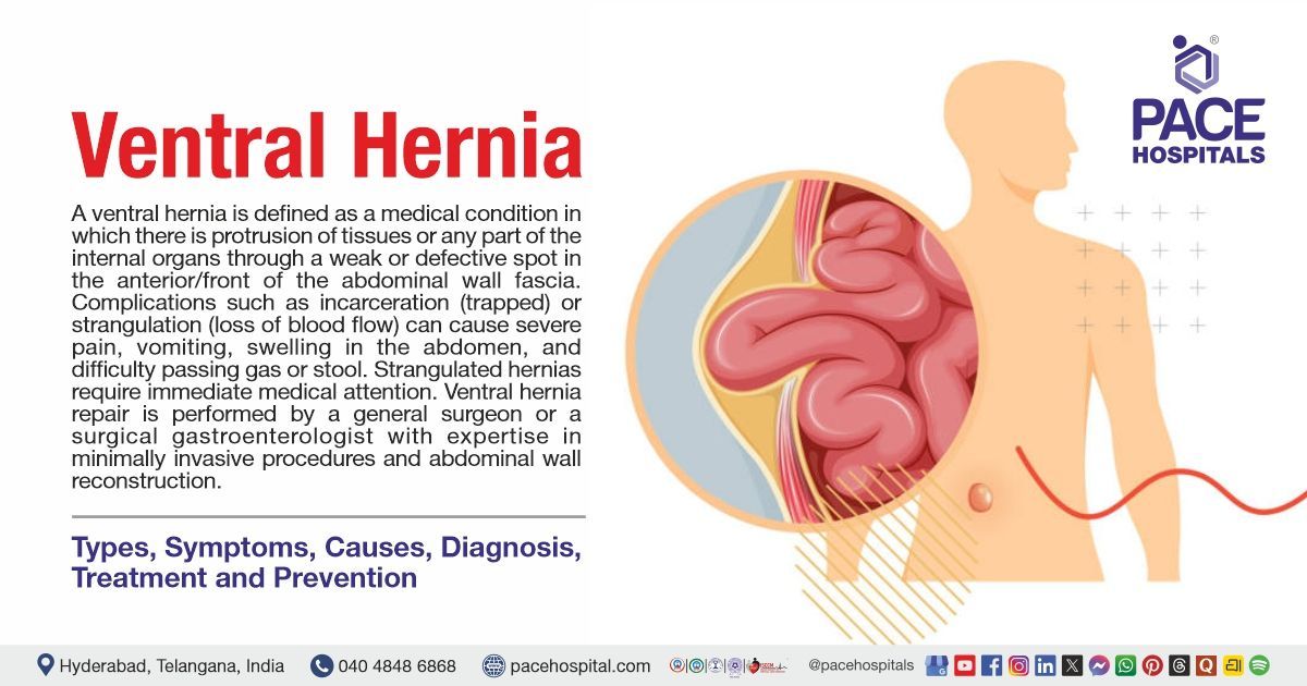
A ventral hernia is defined as a medical condition in which there is protrusion of tissues or any part of the internal organs through a weak or defective spot in the anterior/front of the abdominal wall fascia. It is a broad term that includes primary abdominal-wall hernias, such as umbilical and epigastric hernias, as well as incisional hernias that develop through a scar from a previous abdominal operation.
A ventral hernia may appear as a lump or swelling in the abdomen and can cause pain, pressure, discomfort, or tenderness. The bulge may become more noticeable when a person is standing, coughing, straining, or lifting. Ventral hernias can develop because of weakness in the abdominal wall, including weakness at the site of a previous surgical incision. Factors such as obesity, chronic cough, constipation, pregnancy, and activities that increase pressure inside the abdomen may contribute to hernia development or worsening.
Doctors can usually diagnose a ventral hernia during a physical examination. Ultrasound or CT scan may be advised when the diagnosis is unclear or when a detailed assessment is needed for a large, recurrent, or complex hernia.
Ventral hernia repair is performed by a general surgeon or a specialized surgical gastroenterologist with expertise in abdominal wall reconstruction and minimally invasive or laparoscopic ventral hernia repair techniques.
Ventral hernia meaning
The term ventral hernia originates from Latin words:
- “Ventral” comes from the Latin word venter, meaning “belly” or “abdomen.”
- “Hernia” comes from the Latin word hernia, referring to a protrusion of an organ or tissue through an abnormal opening or weak area.
Thus, a ventral hernia means a protrusion of abdominal tissue or an organ through a weak or defective area of the abdominal wall fascia, usually appearing as a bulge on the front of the abdomen.
Ventral Hernia Prevalence
Ventral hernia prevalence Worldwide
It is estimated that 5% of the general population worldwide has a ventral hernia. Approximately 20% of individuals have primary ventral hernias (such as umbilical or epigastric), whereas 11% to 20% (and up to 30%) of previous midline abdominal surgical incisions result in incisional hernias. Growing risk factors like obesity, ageing populations, and rising rates of abdominal surgery are all contributing to the global burden.
Ventral hernia prevalence in India
In India, abdominal wall and ventral hernias account for 1 to 2 out of every 1,000 patients who are hospitalized for general surgery. Hospital-based research indicates that 4% of all surgical ward admissions are due to ventral hernias, with a significantly higher prevalence in middle-aged and older patients.
Ventral Hernia Types
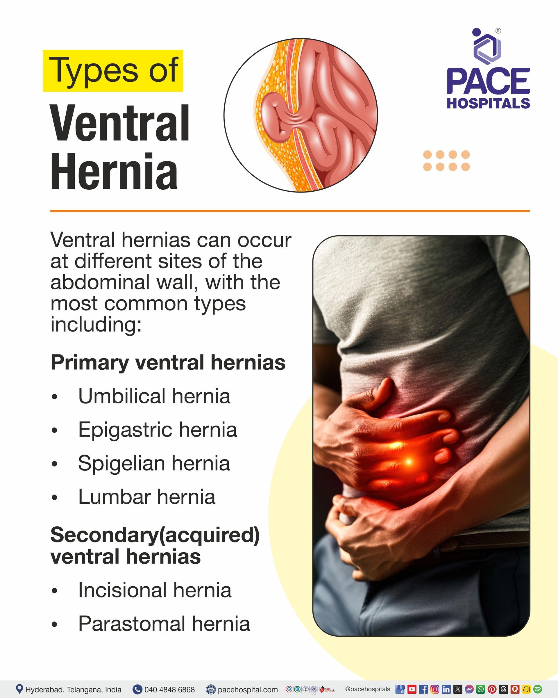
Ventral hernias can occur at different sites of the abdominal wall, with the most common types including:
- Primary ventral hernias
- Umbilical hernia
- Epigastric hernia
- Spigelian hernia
- Lumbar hernia
- Secondary(acquired) ventral hernias
- Incisional hernia
- Parastomal hernia
Primary Ventral Hernias
- Umbilical hernia: An umbilical hernia occurs when a portion of an organ or tissue inside the abdomen pushes out through a weakness in the abdominal area near the belly button. An umbilical hernia is commonly described as the most common primary ventral hernia. In adults, it may develop from high intra-abdominal pressure or abdominal weakness.
- Epigastric hernia: A weakening in the upper midline of the abdominal wall, between the umbilicus and the breastbone (xiphoid process), causes an epigastric hernia. Preperitoneal fat frequently protrudes through a linea alba defect. A visible or palpable bulge and discomfort may result from certain epigastric hernias.
- Spigelian hernia: A defect in the Spigelian fascia along the semilunar line, typically lateral to the rectus abdominis muscle, develops an uncommon type of ventral hernia known as a Spigelian hernia. The lowest portion of the abdominal wall is where it most frequently occurs. Because the protrusion may develop between layers of the abdominal wall, these hernias may be challenging to identify clinically.
- Lumbar hernia:
A lumbar hernia is a rare abdominal wall hernia in which abdominal contents protrude through a weakness or defect in the posterior/lateral abdominal wall. It commonly occurs through the superior (Grynfeltt–Lesshaft) or inferior (Petit) lumbar triangle.
Secondary(acquired) Ventral Hernias
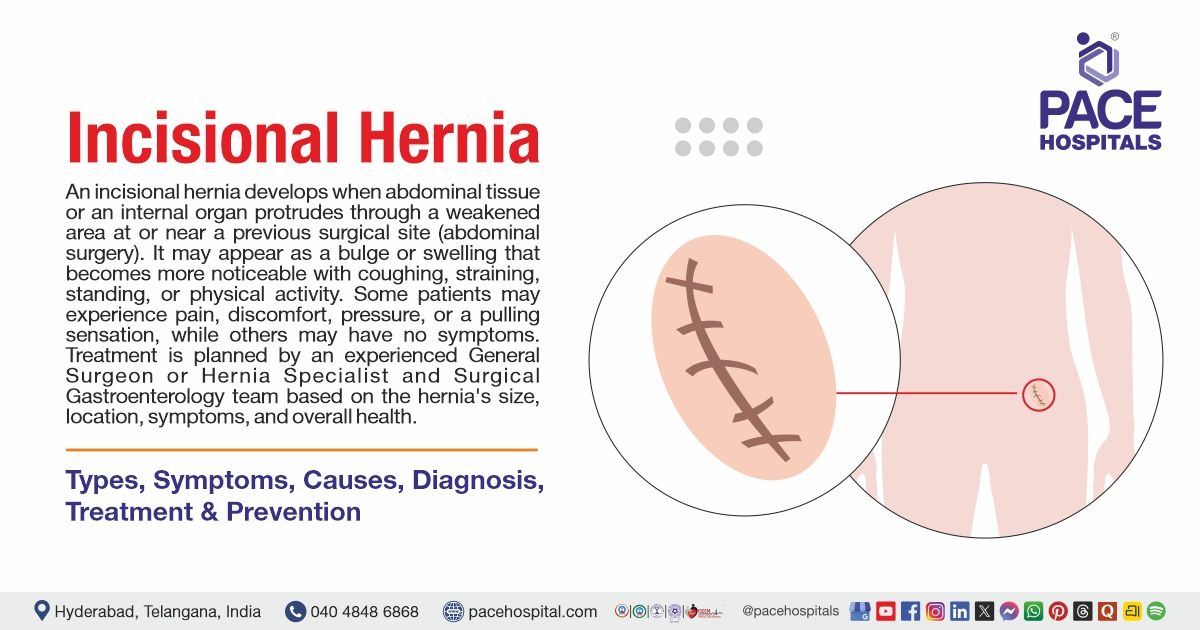
- Incisional hernia: An incisional hernia occurs because of a weak point in the abdomen at the previous operation incision point. The incisional hernia appears as a lump along the incision scar and may appear months or even years after the surgical operation. The defect may allow abdominal tissue or organs to protrude through the weakened area.
- Parastomal hernia: One type of incisional hernia that occurs at or near a stoma, such as an ileostomy or colostomy, is called a parastomal hernia. It happens when an abdominal wall defect formed during stoma development allows abdominal contents to protrude. It can occasionally result in discomfort or other issues, as well as a bulge around the stoma.
Ventral Hernia Symptoms
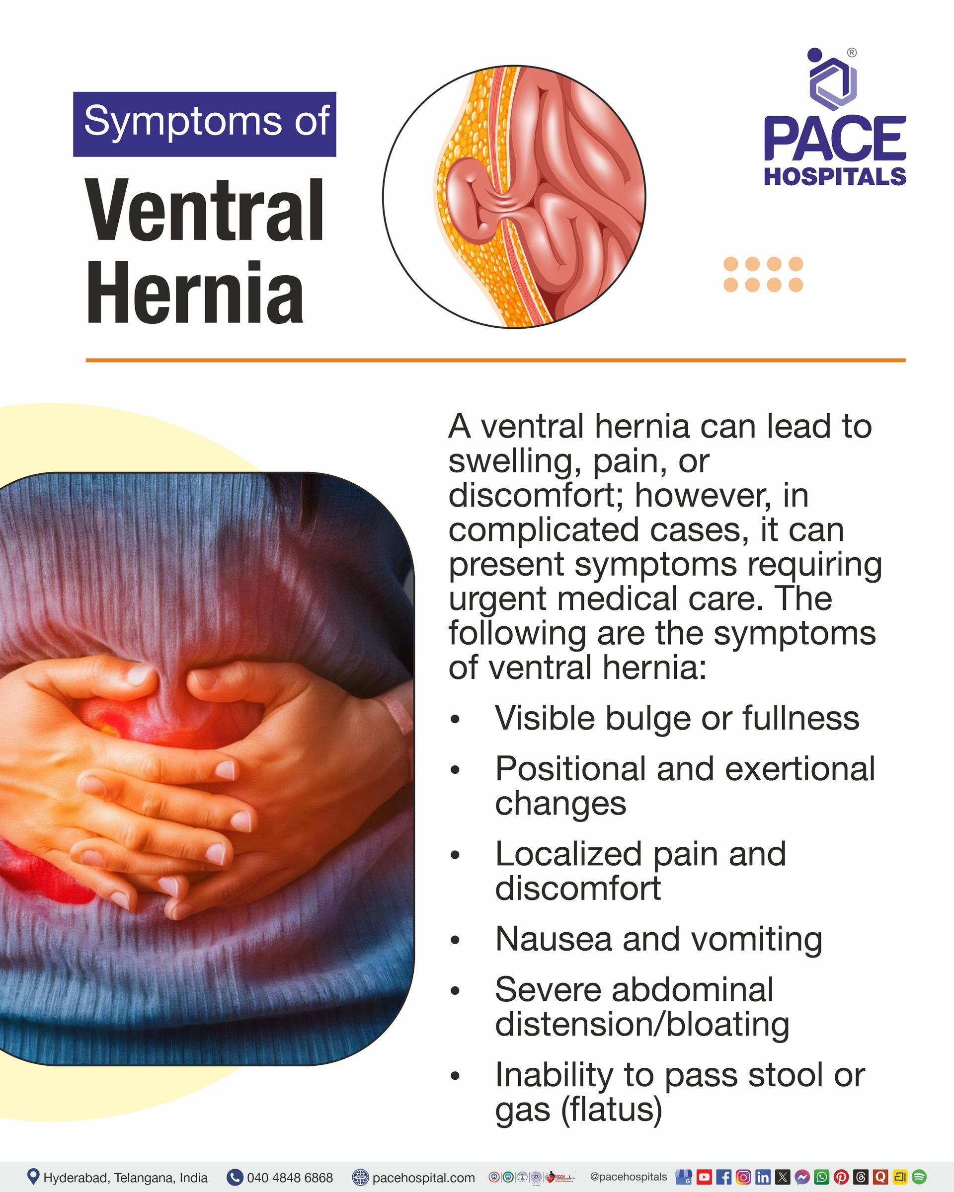
A ventral hernia can lead to swelling, pain, or discomfort; however, in complicated cases, it can present symptoms requiring urgent medical care.
The following are the symptoms of ventral hernia:
- Common/uncomplicated symptoms
- Visible bulge or fullness
- Positional and exertional changes
- Localized pain and discomfort
- Abdominal wall asymmetry
- Complicated/emergency symptoms
- Irreducible bulge
- Severe, acute pain
- Skin discoloration
- Nausea and vomiting
- Severe abdominal distension/bloating
- Inability to pass stool or gas (flatus)
- Systemic signs
Common/uncomplicated Symtoms
Visible bulge or fullness
A soft or swollen area may develop in the abdominal wall and is more noticeable when standing, coughing, straining, or lifting.
Positional and exertional changes
This bulge is more evident when the individual is physically active or standing, and it decreases or disappears when lying down or resting.
Localized pain and discomfort
When coughing, bending, lifting, or engaging in other activities that increase abdominal pressure, some people may experience pain, pressure, tenderness, or discomfort around the hernia.
Abdominal wall asymmetry
One section of the abdominal wall may appear more prominent than the surrounding abdominal surface due to the hernia.
Complicated/Emergency Symptoms
Irreducible bulge
The hernia bulge will be trapped and cannot return to the abdominal cavity, especially if it was previously reducible. This suggests that it is incarcerated and needs medical evaluation.
Severe, acute pain
When the trapped tissue is compromised, especially after strangulation, there may be sudden, intense pain or soreness surrounding the hernia.
Skin discoloration
A reduced blood supply to the trapped tissue may be indicated by the skin over the hernia turning red, purple, blue, or dark.
Nausea and vomiting
These symptoms may occur if the hernia obstructs or compresses the intestines, causing bowel obstruction and preventing normal passage of intestinal contents.
Severe abdominal distension/bloating
When bowel obstruction prevents the normal passage of intestinal contents, it can result in abdominal swelling/bloating.
Inability to pass stool or gas (flatus)
An intestinal blockage linked to a complex hernia may be indicated by the inability to pass gas or bowel movements.
Systemic signs
When strangulation results in tissue damage, infection, or other severe problems, fever and other symptoms of systemic sickness may manifest. These symptoms call for an immediate medical assessment
Causes & Risk Factors of Ventral Hernia
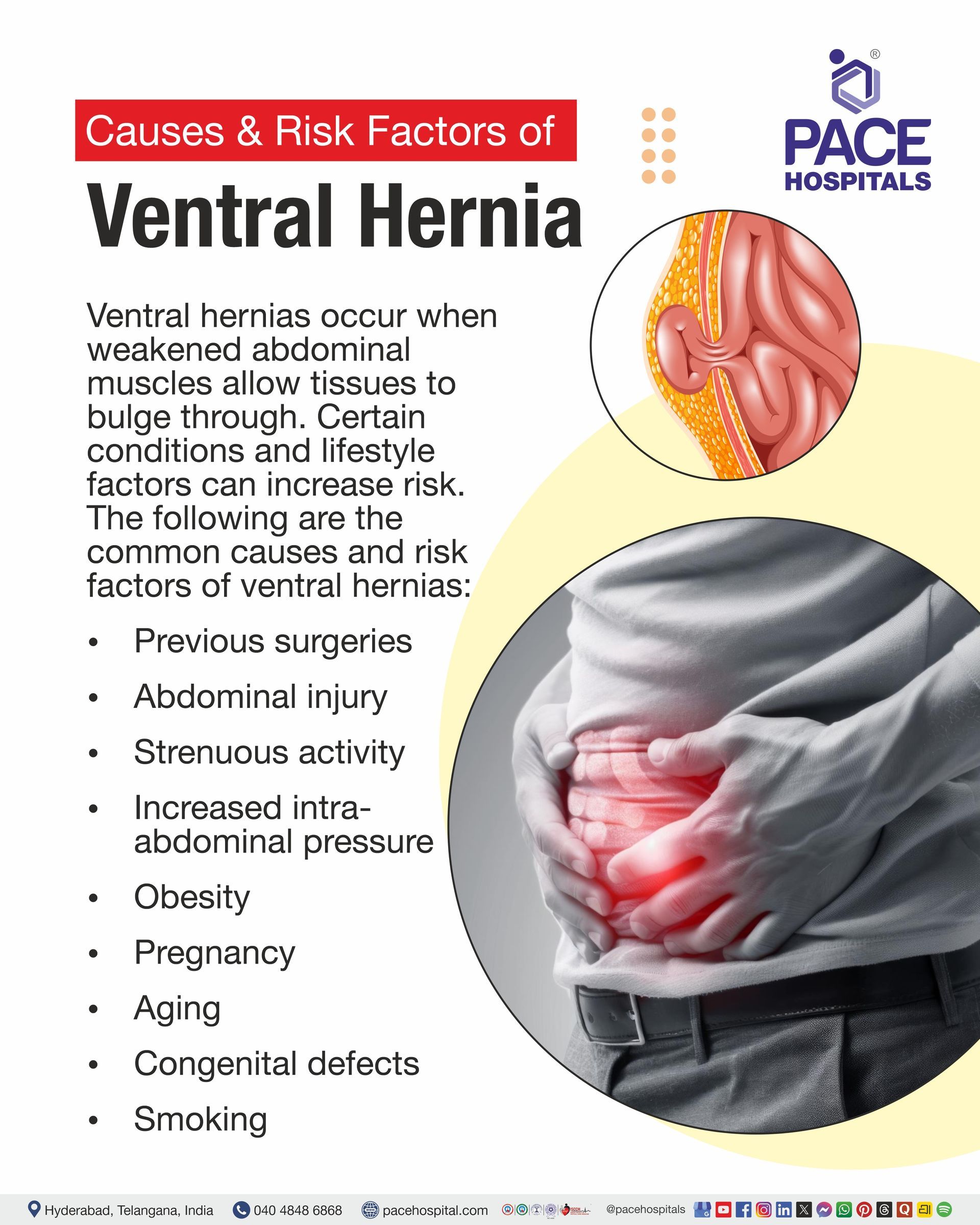
Ventral hernias can occur when weakness in the abdominal wall allows abdominal tissues to bulge through, and certain conditions or lifestyle factors can increase the risk of developing them. The following are the ventral hernia causes and risk factors:
- Previous surgeries
- Abdominal injury
- Strenuous activity
- Increased intra-abdominal pressure
- Obesity
- Pregnancy
- Aging
- Congenital defects
- Smoking
Previous surgeries
Surgical procedures performed on the abdomen can lead to the creation of a weakened portion of the abdominal wall. A hernia that occurs around a past incision area is referred to as an incisional hernia. Weakening of the abdominal wall can persist even after the procedure due to potential infections or other complications during recovery.
Abdominal injury
An injury to the abdominal wall can weaken it by damaging the muscles and connective tissue, allowing abdominal contents to protrude through this weakened region and form a ventral hernia.
Strenuous activity
When there is continuous strain on the abdominal wall from heavy work or other physical activities, there is a greater likelihood of developing a hernia in areas with pre-existing weakness. Strenuous activity may precipitate or make a hernia more noticeable in an area of pre-existing abdominal-wall weakness.
Increased intra-abdominal pressure
Continuous strain on the abdominal wall from increased intra-abdominal pressure can lead to hernia development. Examples of conditions that lead to an increase in intra-abdominal pressure include constipation, chronic cough, vomiting, lifting heavy loads, and childbirth.
Excess weight puts additional stress on the abdominal wall. Obesity increases stress on the abdominal wall and is an established risk factor for ventral hernia.
Pregnancy
During pregnancy, the abdomen becomes larger, which may increase pressure and stretch on the abdominal wall. A ventral wall hernia can be caused by such additional tension.
Aging
With time, the condition of the abdominal wall and its connective tissue may worsen. Ageing may contribute to reduced tissue strength and is a recognized risk factor for ventral hernia.
Congenital defects
Some ventral hernias are congenital, meaning a weakness or defect in the abdominal wall is present from birth. Such hernias may remain unnoticed for years before becoming apparent.
Smoking
Smoking has a detrimental effect on wound healing and tissue quality. Additionally, smoking-related chronic coughing can frequently raise intra-abdominal pressure. As a result, smoking has a special bearing on the development and complications of abdominal wall hernias, particularly following surgery.
Ventral Hernia Complications
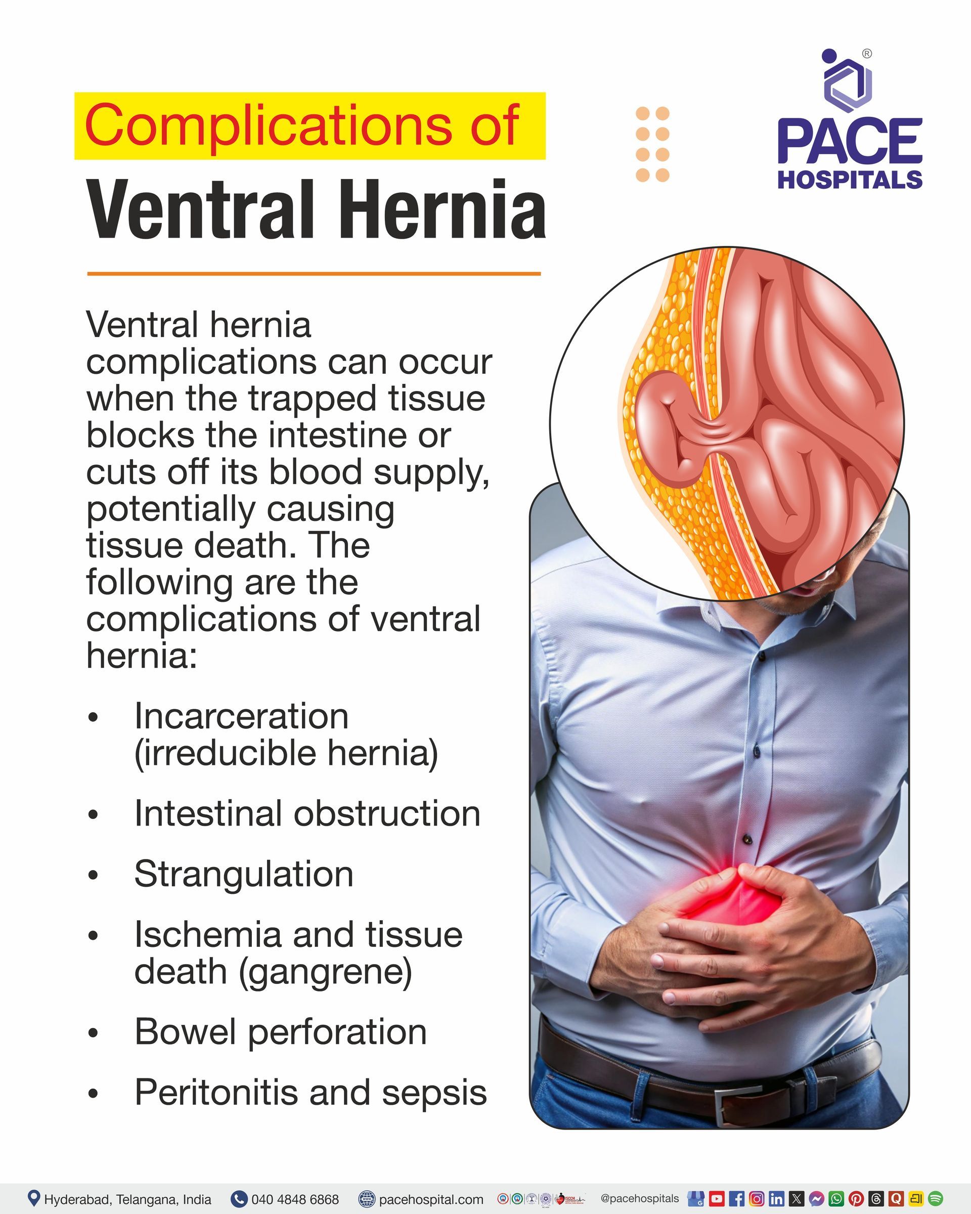
Ventral hernia complications are potentially life-threatening complications resulting from the entrapment of the protruding hernia, which blocks the intestines or stops the flow of blood in them, causing necrosis/tissue death. The following are the complications of ventral hernia:
- Incarceration (irreducible hernia)
- Intestinal obstruction
- Strangulation
- Ischemia and tissue death (gangrene)
- Bowel perforation
- Peritonitis and sepsis
Incarceration (irreducible hernia)
An incarcerated hernia occurs when hernia contents become trapped outside the abdominal wall and cannot be pushed back into the abdomen. It may cause a persistent bulge, discomfort, tenderness, or pain. Incarceration does not always mean that the blood supply has been affected, but it requires prompt medical assessment, especially if the bulge is new, painful, or increasing in size.
Intestinal obstruction
If a portion of the intestine is trapped inside the hernia, the normal passage of intestinal contents may be impeded. This may cause nausea, vomiting, bloating or swelling, difficulty passing gas or stool, and abdominal pain.
Strangulation
This happens when an incarcerated hernia gets so tightly confined that its blood supply is cut off. Because a prolonged reduction of blood flow might result in the death of the damaged intestinal tissue, it is a potentially life-threatening emergency.
Ischemia and tissue death (gangrene)
Ischemia occurs when blood flow is reduced or completely stopped, depriving the affected bowel of oxygen. The damaged bowel may need to be removed if blood flow is not quickly restored because the affected bowel may die or become necrotic.
Bowel perforation
Prolonged strangulation and severe obstruction can harm the intestinal wall and cause a hole or tear in the bowel. A bowel perforation caused by dead or strangled intestines can let pathogens and intestinal contents enter the abdominal cavity.
Peritonitis and sepsis
Peritonitis, a serious inflammation or infection of the abdominal lining, can occur when a perforated or seriously damaged bowel permits bacteria and intestinal contents to enter the abdominal cavity. The illness could then be fatal if the infection spreads throughout the body and causes sepsis.
Note: Seek emergency medical care immediately for a painful hernia that cannot be pushed back, particularly if it is associated with persistent vomiting, abdominal swelling, fever, skin colour changes over the bulge, or worsening pain.
Ventral Hernia Diagnosis
In diagnosing a ventral hernia, the abdominal wall defect is identified by carefully evaluating symptoms and performing a physical examination, with imaging studies as needed.
The following are the steps involved in the diagnosis of a ventral hernia:
- Medical history and symptom evaluation
- Symptom review
- History check
- Complication screen
- Physical examination
- Visual inspection
- Dynamic testing
- Palpation and reducibility
- Ventral hernia diagnostic tests
- Ultrasonography (USG)
- Computed tomography (CT scan)
Medical History and Symptom Evaluation
- Symptom review: The physician will inquire about abdominal distension or protrusion, pain, and exacerbation of symptoms with coughing, straining, lifting, or standing.
- History check: The physician will review any prior abdominal surgeries or other conditions that may contribute to a hernia.
- Complication screen: The physician looks for warning signs, including an abrupt increase or an irreducible bulge, intense pain, redness, fever, nausea, vomiting, or symptoms of intestinal obstruction.
Physical Examination
- Visual inspection: The abdomen is examined for any protrusion or swelling; this may also be assessed by noting any alterations during standing, coughing, or straining.
- Dynamic testing: The patient may be asked to stand, cough, or strain while the physician watches for any bulging of the abdominal wall.
- Palpation and reducibility: The physician then palpates the swelling for tenderness and may attempt to gently move the hernia contents back into the abdominal cavity to assess reducibility.
Ventral hernia diagnostic tests
- Ultrasonography: This may be performed when the diagnosis is unclear after physical examination; it uses sound waves to create images of the abdomen.
- Computed Tomography (CT): CT uses X-rays and a computer to create detailed images that help diagnose the hernia and its complications.
Ventral Hernia Treatment
Ventral hernia treatment depends on the hernia’s size, symptoms, location, and risk of complications. The following are the different treatment options for ventral hernia:
- Surgical management
- Open ventral hernia surgery
- Laparoscopic or robotic ventral hernia repair
- Ventral hernia repair with mesh
- Non-surgical management
- Watchful waiting
- Supportive binders
Surgical Management
- Open ventral hernia surgery: Here, the doctor makes a large incision in the abdomen to directly access, push aside, and repair the weakened abdominal muscles, often using mesh for extra support.
- Laparoscopic or robotic ventral hernia repair: The surgeon makes a few tiny incisions and uses a small camera and specialized instruments to repair the hernia from the inside, often leading to less pain and a faster recovery.
- Ventral hernia repair with mesh:
Mesh is commonly used; synthetic mesh is generally preferred in clean cases, while biologic mesh is reserved for selected situations.
Non-Surgical Management
- Watchful waiting: In cases where the hernia is very small or asymptomatic, the doctor may choose observation rather than immediate surgery, especially when other risk factors are high.
- Supportive binders: An abdominal binder can be used to provide symptomatic relief before surgery, although it will not fix the muscle defect.
Ventral Hernia Prevention
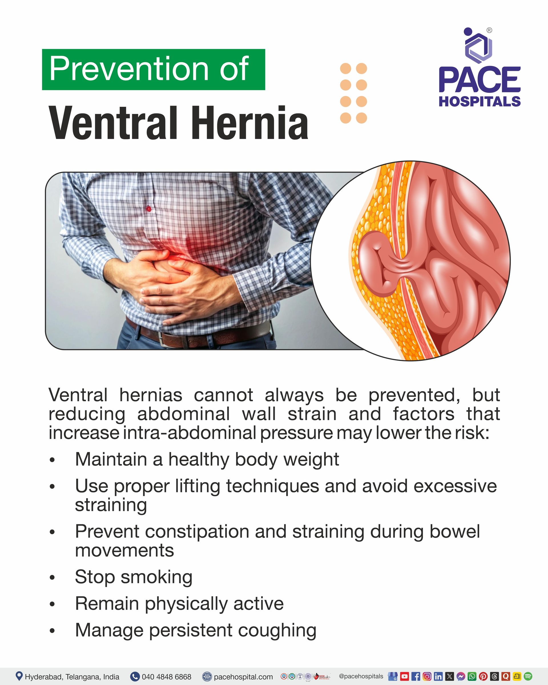
Ventral hernias cannot always be prevented, particularly those related to congenital weakness or previous abdominal surgery. Ventral hernia prevention focuses on reducing factors that weaken the abdominal wall and increase intra-abdominal pressure. The following measures are commonly recommended to reduce the risk of developing a ventral hernia:
- Maintain a healthy body weight
- Use proper lifting techniques and avoid excessive straining
- Prevent constipation and straining during bowel movements
- Stop smoking
- Remain physically active
- Manage persistent coughing
Maintain a healthy body weight
Having a healthy body mass index (BMI) could prevent excess stress and pressure on the abdominal wall. Being overweight is one of the factors increasing the risk of developing a ventral hernia, especially an abdominal-wall hernia and an incisional hernia. If individuals have excess body weight, losing weight slowly through a balanced diet and exercise could reduce their risk.
Use proper lifting techniques and avoid excessive straining
Lifting heavy objects and any activity that significantly increases pressure in the abdominal cavity will add extra stress on the abdominal wall. Therefore, lift properly and do not strain excessively. This applies especially after recent abdominal surgery, when the abdominal wall may still be healing.
Prevent constipation and straining during bowel movements
Abdominal pressure might rise because of persistent constipation and frequent straining to pass stool. Eat enough fiber, stay hydrated, react quickly to the need for a bowel movement, and engage in regular exercise to help prevent constipation. By doing these steps, bowel movements may require less severe straining.
Stop smoking
Smoking can hinder the healing of abdominal surgery wounds and is linked to an increased risk of hernias. Therefore, stopping smoking may enhance wound healing and may lower the chance of an incisional hernia following abdominal surgery. For suitable assistance in quitting smoking, smokers might consult a healthcare provider.
Remain physically active
Staying active is beneficial for maintaining appropriate body weight and general physical fitness. Nevertheless, the exercises must be suitable for the individual's physical condition, and, especially after the abdominal operation, one needs to avoid excessive abdominal pressure and heavy lifting.
Manage persistent coughing
Chronic coughing can increase intra-abdominal pressure, which might strain the abdominal wall. Thus, chronic or recurrent coughs should be managed by the healthcare team, particularly for those with weak abdominal walls or a history of abdominal surgery.
Difference Between Ventral Hernia And Umbilical Hernia
Ventral vs Umbilical hernia
Ventral hernia and umbilical hernia are related but differ in their location, causes, and specific characteristics. Below are the key differences between ventral hernia and umbilical hernia:
| Characteristics | Ventral Hernia | Umbilical Hernia |
|---|---|---|
| Definition | A ventral hernia is a bulge through a weakened area of the abdominal wall. | An umbilical hernia is a bulge that occurs at or around the belly button (umbilicus). |
| Location | It can occur at different sites of the abdominal wall, including areas of previous surgical incisions. | Specifically occurs around the belly button. |
| Common Causes/Risk Factors | May be associated with diabetes, obesity, pregnancy, heavy lifting, coughing, straining, and prior abdominal surgery. | May be caused by the incomplete closure of the abdominal wall around the umbilicus after birth; in adults, pregnancy and excess weight are more common causes. |
| Age Group | Commonly occurs in adults. | Common in infants and adults. |
| Typical appearance | Appears as a bulge in the abdominal wall and may become more prominent with straining. | Usually appears as a gentle lump or soft swelling at the belly button, which can become more prominent with crying, coughing, or straining. |
| Relationship | Ventral hernia is a broad category of hernia that occurs in the abdominal wall. | Umbilical hernia is a specific type of abdominal wall hernia occurring at the umbilicus. |
| Treatment | While complex or symptomatic hernias may need surgical treatment, small, asymptomatic hernias may occasionally be monitored. | Many pediatric umbilical hernias heal on their own; adult hernias that are small and asymptomatic may be monitored, but those that are painful, growing, incarcerated, or strangulated may need surgery. |
Why Choose PACE Hospitals?
Expert Super Specialist Doctors
Advanced Diagnostics & Treatment
Affordable & Transparent Care
24x7 Emergency & ICU Support
Frequently Asked Questions (FAQs) on Ventral Hernia
What is a ventral hernia?
A ventral hernia is a weakness or opening in the abdominal wall that permits abdominal tissue, such as fatty tissue, small bowel, colon, or omentum, to protrude and form a visible or felt bulge. Ventral hernias can arise in the umbilical, para-umbilical, epigastric, or incisional regions.
What is a ventral hernia repair?
Ventral Hernia Repair is a surgical procedure performed to restore the hernia contents to their original position and strengthen the weakened part of the abdominal wall. According to the type, size and location of the hernia, the operation can either be done using keyhole surgery or traditional surgery. This operation can be done by repairing the defect or weakness in the abdominal wall, using a surgical mesh to reinforce the weakened area, or a combination of both methods.No. A femoral hernia does not usually heal on its own because the underlying weakness or defect in the abdominal wall does not close spontaneously. Symptoms may temporarily improve, but the hernia can persist and may become incarcerated or strangulated. Therefore, medical evaluation and timely surgical repair are recommended for a femoral hernia.
Are ventral hernia and umbilical hernia the same?
No, an umbilical hernia is a ventral hernia that occurs at the belly button. The term ventral hernia is broader and includes umbilical, paraumbilical, epigastric, and incisional hernias, which occur at various sites on the anterior part of the abdominal wall.
What causes ventral hernia?
A ventral hernia can occur when tissue from the abdomen pushes outward due to a weak or damaged abdominal wall. The defect may be present from birth or develop later in life. Pregnancy, obesity, increased abdominal pressure, and previous abdominal surgery are all associated with abdominal wall hernias. The exact cause may vary depending on the type of ventral hernia.
What are the common symptoms of a ventral hernia?
A lump or protrusion on the abdominal wall, which may become more apparent when coughing, straining, or standing, is the most typical indication of a ventral hernia. While some people may not feel any pain at all, others may feel pain, tenderness, or discomfort near the hernia. A medical evaluation is necessary if the hernia becomes painful, hard, swollen, or difficult to reenter the abdomen.
What is the difference between a ventral hernia and an abdominal hernia?
Although the terms can occasionally be used interchangeably, a ventral hernia refers exclusively to a hernia that develops on the front wall of the abdomen. "Abdominal hernia" is a more general term for hernias that develop in the abdomen. Certain types of ventral hernias include incisional, umbilical, para-umbilical, and epigastric hernias. Also, ventral hernias can include lateral defects such as Spigelian hernias.
Can a ventral hernia go away on its own without surgery?
Adult ventral hernias usually do not heal on their own without surgical intervention. Because adult umbilical hernias are unlikely to cure on their own, surgery is usually recommended when necessary. But not all hernias require emergency surgery; the decision depends on the patient's overall health, the type and characteristics of the hernia, and the symptoms.
When does a ventral hernia need surgery?
Surgery may be recommended if a ventral hernia enlarges, becomes problematic, causes significant or persistent pain or discomfort, or interferes with everyday activities. An individual assessment is essential since the type, size, and location of the hernia, along with the patient's symptoms and overall health, decide whether and when surgery is required.
What happens if a ventral hernia is left untreated?
In certain individuals, an untreated ventral hernia may stay stable, but it may also progressively enlarge and become trapped. The tissue within the hernia may occasionally become incarcerated, preventing it from being forced back into the abdomen. Strangulation may happen, and immediate medical attention may be necessary if the blood supply to the confined tissue is cut off.
Can a ventral hernia become dangerous or life-threatening?
Yes, a ventral hernia can become dangerous if its contents become trapped and the blood supply is cut off, even if serious complications are rare. This is a medical emergency known as a strangulated hernia. Severe problems, such as bowel injury, gangrene, and sepsis, can result from strangulation of gut tissue, necessitating immediate surgery.
What is the best treatment for a ventral hernia?
For every ventral hernia, there isn't a single best treatment. The optimal course of treatment depends on the patient's medical history, general health, the type, size, and location of the hernia, as well as the symptoms it produces. If surgery is required, the abdominal wall may be repaired using mesh, sutures, open or keyhole surgery, or a combination of techniques.
What are the different types of ventral hernias?
There are several places on the front of the abdomen where ventral hernias can develop. Umbilical hernias occur at the belly button; para-umbilical hernias occur near the belly button; epigastric hernias occur between the lower breastbone and the belly button; and incisional hernias develop at the site of an earlier abdominal surgical incision.
What is the recovery time after ventral hernia surgery?
Recovery following ventral hernia repair depends on the nature and complexity of the hernia, the surgical procedure performed, and the individual's health status. Recovery from the repair of an umbilical hernia requires 4 to 6 weeks. But patients can gradually resume their usual tasks as their recovery allows and according to the healthcare provider’s recommendations.
How long after ventral hernia surgery can a person return to work?
The type of surgery and the job's physical demands are the primary determinants of the time required to return to work. Many patients can resume their jobs in one to two weeks, but individuals whose jobs require heavy lifting or physically demanding tasks could need more time to recover. Some incisional hernia repair notes that 2 to 4 weeks may be typical, with individual recovery varying from patient to patient.
When can a person exercise or lift weights after ventral hernia surgery?
In general, patients are advised to begin with mild exercise and progressively increase it as they heal. Depending on the type of repair, the timing for resuming heavy lifting and vigorous exercise varies. Heavy lifting and intense exercise should be avoided for many weeks following hernia surgery. Some guidelines suggest waiting four to six weeks after correcting an abdominal or umbilical hernia. Based on the repair and recovery, the surgeon should provide personalized counsel.
What should a person eat after ventral hernia surgery?
Unless their medical team advises otherwise, most people can gradually return to their normal diet following hernia surgery. Drinking enough water and eating foods high in fibre, such as fruits, vegetables, whole grains, and oats, can help prevent constipation. Constipation and extreme straining can put pressure on the healing abdominal wall; therefore, it's very important to avoid them during rehabilitation.
Can a ventral hernia come back after surgery?
Yes. Even with long-lasting hernia repair, recurrence is still possible. The type and size of the hernia, the patient's characteristics, and the method of repair all affect the risk. Recovery might be aided by gradually resuming physical activity following postoperative instructions.
What are the possible complications of ventral hernia surgery?
Most patients will make a full recovery without complications; however, as with any surgical procedure, ventral hernia repair may carry risks. Complications of ventral hernia repair include wound infection, bleeding, hematoma, seroma, persistent pain, blood clots, and recurrence of the hernia. In some instances, internal organs in the abdominal cavity, including intestines and blood vessels, may be damaged by surgical intervention. The specific risks depend on the type and complexity of the repair and should be discussed with the surgeon before the procedure.
When to consult a doctor for ventral hernia?
Consult a doctor for a ventral hernia if you notice a persistent or increasing bulge in the abdominal wall, pain, discomfort, or symptoms that interfere with your daily activities. Signs that indicate a need for medical evaluation are:
- A new or enlarging abdominal bulge
- Persistent pain, tenderness, or discomfort around the hernia
- A bulge that becomes difficult to push back or does not reduce when lying down
- Increasing abdominal swelling or bloating
- Nausea or vomiting associated with the hernia
- Difficulty passing stool or gas
- Symptoms that worsen during physical activity, coughing, or straining
If these symptoms continue or worsen, it is best to see a doctor for an accurate diagnosis and appropriate treatment. Seek emergency medical attention if the hernia becomes suddenly painful, firm, swollen, discoloured, or cannot be pushed back, especially if accompanied by vomiting or inability to pass stool or gas, as these may indicate incarceration, bowel obstruction, or strangulation. A general surgeon or gastroenterologist can assess the condition and refer to a general surgeon when surgical treatment is required.
Share on
Request an appointment
Fill in the appointment form or call us instantly to book a confirmed appointment with our super specialist at 04048486868
Appointment request - health articles
Recent Articles